<?xml version='1.0' encoding='UTF-8'?>

<!DOCTYPE article PUBLIC "-//NLM//DTD JATS (Z39.96) Journal Publishing DTD v1.1d1 20130915//EN" "JATS-journalpublishing1.dtd">
<article xmlns:xlink="http://www.w3.org/1999/xlink">
  <front>
    <journal-meta id="journal-meta-87cddb9ab7774ac9973b6a64b7cbc767">
      <journal-id journal-id-type="nlm-ta">Sciresol</journal-id>
      <journal-id journal-id-type="publisher-id">Sciresol</journal-id>
      <journal-id journal-id-type="journal_submission_guidelines">https://jmsh.ac.in/</journal-id>
      <journal-title-group>
        <journal-title>Journal of Medical Sciences and Health</journal-title>
      </journal-title-group>
      <issn publication-format="print"/>
    </journal-meta>
    <article-meta>
        
          
            <article-id pub-id-type="doi">10.47799/pimr.1402.25.84</article-id>
          
          
            <article-categories>
              <subj-group>
                <subject>REVIEW ARTICLE</subject>
              </subj-group>
            </article-categories>
            <title-group>
              <article-title>&lt;p&gt;Apoptosis in Cancer: From Threat to Therapy&lt;/p&gt;</article-title>
            </title-group>
          
          
            <pub-date date-type="pub">
              <day>30</day>
              <month>3</month>
              <year>2026</year>
            </pub-date>
            <permissions>
              <copyright-year>2026</copyright-year>
            </permissions>
          
          
            <volume>14</volume>
          
          
            <issue>2</issue>
          
          <fpage>1</fpage>

          <abstract>
            <title>Abstract</title>
            &lt;p&gt;Apoptosis is a tightly regulated form of programmed cell death essential for tissue homeostasis and genomic integrity. Its dysregulation contributes to diverse pathological conditions, particularly cancer, where evasion of apoptosis enables malignant transformation, tumour progression, immune escape, and therapeutic resistance. Contemporary understanding recognises apoptosis as an integrated network involving mitochondrial, death receptor, and caspase-dependent signalling pathways rather than isolated linear cascades. This review synthesises current insights into the molecular mechanisms governing apoptotic regulation and examines how disruptions in BCL-2 family proteins, p53 signalling, caspase activation, and inhibitor of apoptosis proteins promote carcinogenesis. Particular emphasis is placed on translational advances in apoptosis-targeted therapy, including the clinical success of BH3 mimetics, emerging p53-modulating strategies, and combinational approaches designed to overcome apoptotic resistance. By integrating mechanistic biology with therapeutic development, this review highlights apoptosis not only as a hallmark of cancer but also as a therapeutically exploitable vulnerability in selected cancer contexts, particularly in haematological malignancies, although its translation in solid tumours remains variable and often limited. Understanding tumour-specific apoptotic dependencies may enable more precise and less toxic anticancer strategies in the evolving era of precision oncology.&lt;/p&gt;
          </abstract>
          
          
            <kwd-group>
              <title>Keywords</title>
              
                <kwd>Apoptosis; BCL-2 family; Caspases; p53; BH3 mimetics; Cancer therapy</kwd>
              
            </kwd-group>


        <contrib-group>
          
            
              <contrib contrib-type="author">
                <name>
                  <surname>Chagalamari</surname>
                  <given-names>Akhila</given-names>
                </name>
                
                  <xref rid="aff-1" ref-type="aff">1</xref>
                
              </contrib>


              <aff id="aff-1">
                <institution> Assistant Professor, Department of Pharmacology Government Medical College </institution>
                <addr-line>Piduguralla, Andhra Pradesh India</addr-line>
              </aff>
            
              <aff id="aff-2">
                <institution> Associate Professor, Department of Pharmacology AIIMS Mangalagiri </institution>
                <addr-line>Andhra Pradesh India</addr-line>
              </aff>
            
              <aff id="aff-3">
                <institution> Assistant Professor of Cardiology &amp; Cardiac Electrophysiology Sri Jayadeva Institute of Cardiovascular Sciences and Research </institution>
                <addr-line>India </addr-line>
              </aff>
            
              <aff id="aff-4">
                <institution> Professor and Head, Department of Pharmacology AIIMS Mangalagiri </institution>
                <addr-line>Andhra Pradesh India</addr-line>
              </aff>
            
              <aff id="aff-5">
                <institution> Additional Professor, Department of Pharmacology AIIMS Mangalagiri </institution>
                <addr-line>Andhra Pradesh India</addr-line>
              </aff>
            
              <aff id="aff-6">
                <institution> Professor, Department of Community Medicine Maheswara Medical College </institution>
                <addr-line>Hyderabad, Telangana India</addr-line>
              </aff>


              <contrib contrib-type="author">
                <name>
                  <surname>Rangari</surname>
                  <given-names>Gaurav M</given-names>
                </name>
                
                  <xref rid="aff-2" ref-type="aff">2</xref>
                
              </contrib>


              <aff id="aff-1">
                <institution> Assistant Professor, Department of Pharmacology Government Medical College </institution>
                <addr-line>Piduguralla, Andhra Pradesh India</addr-line>
              </aff>
            
              <aff id="aff-2">
                <institution> Associate Professor, Department of Pharmacology AIIMS Mangalagiri </institution>
                <addr-line>Andhra Pradesh India</addr-line>
              </aff>
            
              <aff id="aff-3">
                <institution> Assistant Professor of Cardiology &amp; Cardiac Electrophysiology Sri Jayadeva Institute of Cardiovascular Sciences and Research </institution>
                <addr-line>India </addr-line>
              </aff>
            
              <aff id="aff-4">
                <institution> Professor and Head, Department of Pharmacology AIIMS Mangalagiri </institution>
                <addr-line>Andhra Pradesh India</addr-line>
              </aff>
            
              <aff id="aff-5">
                <institution> Additional Professor, Department of Pharmacology AIIMS Mangalagiri </institution>
                <addr-line>Andhra Pradesh India</addr-line>
              </aff>
            
              <aff id="aff-6">
                <institution> Professor, Department of Community Medicine Maheswara Medical College </institution>
                <addr-line>Hyderabad, Telangana India</addr-line>
              </aff>


              <contrib contrib-type="author">
                <name>
                  <surname>Rao</surname>
                  <given-names>Bhaskar S</given-names>
                </name>
                
                  <xref rid="aff-3" ref-type="aff">3</xref>
                
              </contrib>


              <aff id="aff-1">
                <institution> Assistant Professor, Department of Pharmacology Government Medical College </institution>
                <addr-line>Piduguralla, Andhra Pradesh India</addr-line>
              </aff>
            
              <aff id="aff-2">
                <institution> Associate Professor, Department of Pharmacology AIIMS Mangalagiri </institution>
                <addr-line>Andhra Pradesh India</addr-line>
              </aff>
            
              <aff id="aff-3">
                <institution> Assistant Professor of Cardiology &amp; Cardiac Electrophysiology Sri Jayadeva Institute of Cardiovascular Sciences and Research </institution>
                <addr-line>India </addr-line>
              </aff>
            
              <aff id="aff-4">
                <institution> Professor and Head, Department of Pharmacology AIIMS Mangalagiri </institution>
                <addr-line>Andhra Pradesh India</addr-line>
              </aff>
            
              <aff id="aff-5">
                <institution> Additional Professor, Department of Pharmacology AIIMS Mangalagiri </institution>
                <addr-line>Andhra Pradesh India</addr-line>
              </aff>
            
              <aff id="aff-6">
                <institution> Professor, Department of Community Medicine Maheswara Medical College </institution>
                <addr-line>Hyderabad, Telangana India</addr-line>
              </aff>


              <contrib contrib-type="author">
                <name>
                  <surname>Sharma</surname>
                  <given-names>Sushil</given-names>
                </name>
                
                  <xref rid="aff-4" ref-type="aff">4</xref>
                
              </contrib>


              <aff id="aff-1">
                <institution> Assistant Professor, Department of Pharmacology Government Medical College </institution>
                <addr-line>Piduguralla, Andhra Pradesh India</addr-line>
              </aff>
            
              <aff id="aff-2">
                <institution> Associate Professor, Department of Pharmacology AIIMS Mangalagiri </institution>
                <addr-line>Andhra Pradesh India</addr-line>
              </aff>
            
              <aff id="aff-3">
                <institution> Assistant Professor of Cardiology &amp; Cardiac Electrophysiology Sri Jayadeva Institute of Cardiovascular Sciences and Research </institution>
                <addr-line>India </addr-line>
              </aff>
            
              <aff id="aff-4">
                <institution> Professor and Head, Department of Pharmacology AIIMS Mangalagiri </institution>
                <addr-line>Andhra Pradesh India</addr-line>
              </aff>
            
              <aff id="aff-5">
                <institution> Additional Professor, Department of Pharmacology AIIMS Mangalagiri </institution>
                <addr-line>Andhra Pradesh India</addr-line>
              </aff>
            
              <aff id="aff-6">
                <institution> Professor, Department of Community Medicine Maheswara Medical College </institution>
                <addr-line>Hyderabad, Telangana India</addr-line>
              </aff>


              <contrib contrib-type="author">
                <name>
                  <surname></surname>
                  <given-names>Madhavrao</given-names>
                </name>
                
                  <xref rid="aff-5" ref-type="aff">5</xref>
                
              </contrib>


              <aff id="aff-1">
                <institution> Assistant Professor, Department of Pharmacology Government Medical College </institution>
                <addr-line>Piduguralla, Andhra Pradesh India</addr-line>
              </aff>
            
              <aff id="aff-2">
                <institution> Associate Professor, Department of Pharmacology AIIMS Mangalagiri </institution>
                <addr-line>Andhra Pradesh India</addr-line>
              </aff>
            
              <aff id="aff-3">
                <institution> Assistant Professor of Cardiology &amp; Cardiac Electrophysiology Sri Jayadeva Institute of Cardiovascular Sciences and Research </institution>
                <addr-line>India </addr-line>
              </aff>
            
              <aff id="aff-4">
                <institution> Professor and Head, Department of Pharmacology AIIMS Mangalagiri </institution>
                <addr-line>Andhra Pradesh India</addr-line>
              </aff>
            
              <aff id="aff-5">
                <institution> Additional Professor, Department of Pharmacology AIIMS Mangalagiri </institution>
                <addr-line>Andhra Pradesh India</addr-line>
              </aff>
            
              <aff id="aff-6">
                <institution> Professor, Department of Community Medicine Maheswara Medical College </institution>
                <addr-line>Hyderabad, Telangana India</addr-line>
              </aff>


              <contrib contrib-type="author">
                <name>
                  <surname>Misra</surname>
                  <given-names>Arup Kumar</given-names>
                </name>
                
                  <xref rid="aff-2" ref-type="aff">2</xref>
                
              </contrib>


              <aff id="aff-1">
                <institution> Assistant Professor, Department of Pharmacology Government Medical College </institution>
                <addr-line>Piduguralla, Andhra Pradesh India</addr-line>
              </aff>
            
              <aff id="aff-2">
                <institution> Associate Professor, Department of Pharmacology AIIMS Mangalagiri </institution>
                <addr-line>Andhra Pradesh India</addr-line>
              </aff>
            
              <aff id="aff-3">
                <institution> Assistant Professor of Cardiology &amp; Cardiac Electrophysiology Sri Jayadeva Institute of Cardiovascular Sciences and Research </institution>
                <addr-line>India </addr-line>
              </aff>
            
              <aff id="aff-4">
                <institution> Professor and Head, Department of Pharmacology AIIMS Mangalagiri </institution>
                <addr-line>Andhra Pradesh India</addr-line>
              </aff>
            
              <aff id="aff-5">
                <institution> Additional Professor, Department of Pharmacology AIIMS Mangalagiri </institution>
                <addr-line>Andhra Pradesh India</addr-line>
              </aff>
            
              <aff id="aff-6">
                <institution> Professor, Department of Community Medicine Maheswara Medical College </institution>
                <addr-line>Hyderabad, Telangana India</addr-line>
              </aff>


              <contrib contrib-type="author">
                <name>
                  <surname>Bhaisare</surname>
                  <given-names>Roza</given-names>
                </name>
                
                  <xref rid="aff-6" ref-type="aff">6</xref>
                
              </contrib>


              <aff id="aff-1">
                <institution> Assistant Professor, Department of Pharmacology Government Medical College </institution>
                <addr-line>Piduguralla, Andhra Pradesh India</addr-line>
              </aff>
            
              <aff id="aff-2">
                <institution> Associate Professor, Department of Pharmacology AIIMS Mangalagiri </institution>
                <addr-line>Andhra Pradesh India</addr-line>
              </aff>
            
              <aff id="aff-3">
                <institution> Assistant Professor of Cardiology &amp; Cardiac Electrophysiology Sri Jayadeva Institute of Cardiovascular Sciences and Research </institution>
                <addr-line>India </addr-line>
              </aff>
            
              <aff id="aff-4">
                <institution> Professor and Head, Department of Pharmacology AIIMS Mangalagiri </institution>
                <addr-line>Andhra Pradesh India</addr-line>
              </aff>
            
              <aff id="aff-5">
                <institution> Additional Professor, Department of Pharmacology AIIMS Mangalagiri </institution>
                <addr-line>Andhra Pradesh India</addr-line>
              </aff>
            
              <aff id="aff-6">
                <institution> Professor, Department of Community Medicine Maheswara Medical College </institution>
                <addr-line>Hyderabad, Telangana India</addr-line>
              </aff>
            
          
        </contrib-group>
        
    </article-meta>
  </front>
  <body>
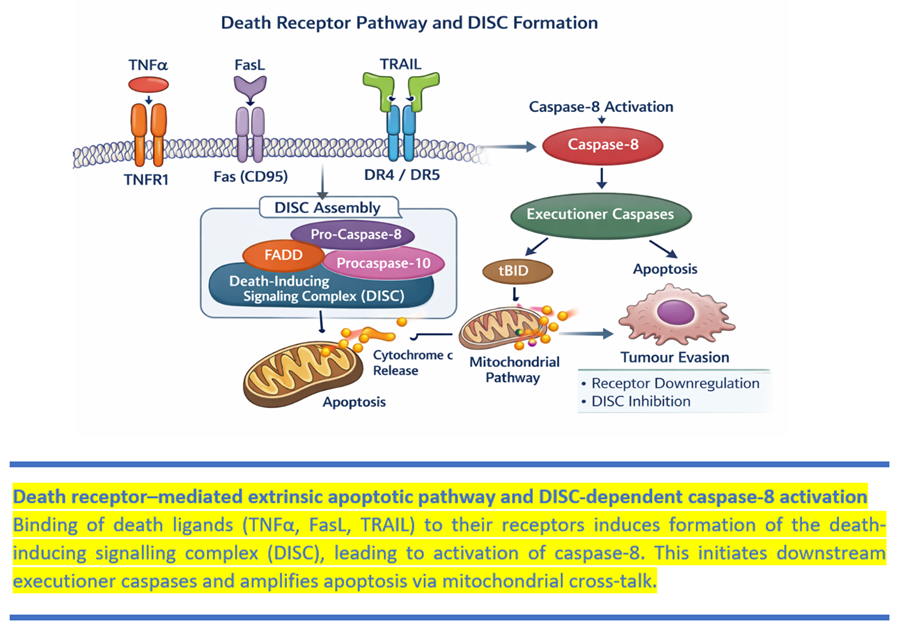
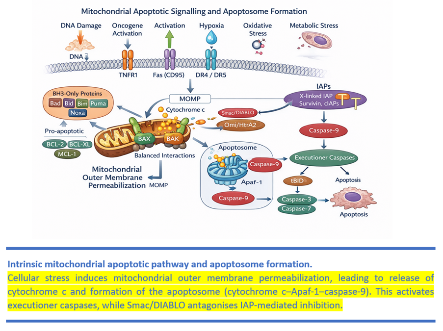
    <heading><span><bold>INTRODUCTION </bold></span></heading><p><span>Apoptosis is a regulated, energy-dependent process essential for maintaining tissue homeostasis and genomic stability. It plays critical roles in development, immune regulation, and elimination of damaged or potentially malignant cells. Dysregulation of apoptosis contributes to multiple pathological conditions, most notably cancer, where evasion of programmed cell death enables tumour survival, progression, and resistance to therapy<superscript>[<xref ref-type="link" rid="#ref-1">1</xref>-<xref ref-type="link" rid="#ref-3">3</xref>]</superscript>.</span></p><p><span>While apoptosis has historically been described as a linear cascade, contemporary evidence supports a network-based model involving interconnected mitochondrial, death receptor, and caspase-mediated pathways<superscript>[<xref ref-type="link" rid="#ref-4">4</xref>, <xref ref-type="link" rid="#ref-5">5</xref>]</superscript>. These pathways are tightly regulated and function as a critical barrier against oncogenesis.</span></p><p><span>In cancer, disruption of apoptotic signalling occurs through alterations in BCL-2 family proteins, p53 dysfunction, impaired caspase activation, and overexpression of inhibitor of apoptosis proteins (IAPs). These changes collectively enable tumour cells to evade cell death despite genomic instability and therapeutic stress<superscript>[<xref ref-type="link" rid="#ref-6">6</xref>-<xref ref-type="link" rid="#ref-8">8</xref>]</superscript>.</span></p><p><span>Therapeutic targeting of apoptosis has gained considerable attention, particularly with the development of BH3 mimetics such as venetoclax, which have shown clinical success in haematological malignancies. However, translation into solid tumours remains challenging due to tumour heterogeneity, adaptive resistance, and redundancy in survival pathways<superscript>[<xref ref-type="link" rid="#ref-9">9</xref>-<xref ref-type="link" rid="#ref-11">11</xref>]</superscript>.</span></p><p><span>This review provides a concise, critically synthesised overview of apoptotic mechanisms, their role in carcinogenesis, and current therapeutic strategies, with particular emphasis on clinical limitations and future directions. </span></p><heading><span><bold>MORPHOLOGICAL AND BIOCHEMICAL FEATURES OF APOPTOSIS</bold></span></heading><p><span>Apoptosis is defined not only by its molecular regulation but also by its distinct structural and biochemical characteristics, which differentiate it from necrosis and other forms of regulated cell death. These features are critical for diagnostic recognition and for understanding how apoptotic signalling translates into controlled cellular dismantling.</span></p><p><span>Unlike necrosis, apoptosis is characterised by preservation of membrane integrity, absence of inflammation, and orderly cellular fragmentation into apoptotic bodies<superscript>[<xref ref-type="link" rid="#ref-1">1</xref>-<xref ref-type="link" rid="#ref-5">5</xref>]</superscript>.</span></p><p><span>These morphological changes are closely coordinated with underlying biochemical processes that ensure orderly cellular dismantling and non-inflammatory clearance. An early and functionally critical biochemical event in apoptosis is the externalisation of phosphatidylserine from the inner to the outer leaflet of the plasma membrane. This process, mediated by coordinated regulation of membrane phospholipid transporters, serves as an “eat-me” signal that facilitates recognition and rapid clearance of apoptotic cells by macrophages<superscript>[<xref ref-type="link" rid="#ref-4">4</xref>]</superscript>.  This mechanism ensures non-inflammatory removal of dying cells and preservation of tissue homeostasis<superscript>[<xref ref-type="link" rid="#ref-6">6</xref>]</superscript>.</span></p><p><span>Markers such as caspase activation or DNA fragmentation are useful but not definitive: apoptosis can occur with limited DNA laddering and, in some contexts, with reduced caspase dependence. Classification should integrate morphology with molecular context rather than rely on a single assay<superscript>[<xref ref-type="link" rid="#ref-2">2</xref>, <xref ref-type="link" rid="#ref-7">7</xref>]</superscript>.</span></p><p><span>Collectively, these morphological and biochemical changes reflect a highly regulated cellular self-destruction program designed to eliminate unwanted cells while preserving tissue integrity. In the context of cancer biology, disruption of these tightly controlled processes enables tumour cell survival and underpins resistance to therapy.</span></p><heading><span><bold>MECHANISMS OF APOPTOSIS</bold></span></heading><p><span>Apoptosis is best understood as an interconnected signalling network rather than a strictly linear pathway, with multiple upstream inputs converging on a common execution phase<superscript>[<xref ref-type="link" rid="#ref-7">7</xref>]</superscript>.</span></p><p><span>Caspases are central mediators of apoptosis and function as a hierarchical proteolytic cascade. Initiator caspases (such as caspase-8 and -9) respond to upstream activation signals and subsequently activate executioner caspases (caspase-3 and -7), which mediate cleavage of key structural and regulatory proteins, leading to the morphological and biochemical features of apoptosis<superscript>[<xref ref-type="link" rid="#ref-8">8</xref>]</superscript>.</span></p><p><italic><span><bold>The Extrinsic (Death Receptor) Pathway</bold></span></italic></p><p><span>The extrinsic apoptotic pathway is initiated at the cell surface following engagement of specific death receptors belonging to the tumour necrosis factor (TNF) receptor superfamily. Key receptors include TNFR1, Fas (CD95), and TRAIL receptors DR4 and DR5. Ligand binding induces receptor trimerization and recruitment of adaptor proteins such as Fas-associated death domain (FADD), resulting in assembly of the death-inducing signalling complex (DISC) <superscript>[<xref ref-type="link" rid="#ref-9">9</xref>]</superscript> <xref ref-type="link" rid="#figure-1">[Fig. 1]</xref>.</span></p><p><span>DISC assembly activates caspase-8, which triggers executioner caspases (caspase-3/-7) and, via BID cleavage to tBID, can amplify death signals through mitochondria. Tumours evade this axis through reduced receptor expression, altered DISC components, or downstream inhibition, limiting immune-mediated killing and TRAIL responses<superscript>[<xref ref-type="link" rid="#ref-9">9</xref>]</superscript>.</span></p><p><span>In cancer biology, defects in the extrinsic pathway-such as reduced death receptor expression, increased decoy receptors, or impaired DISC formation-contribute to resistance against immune-mediated cytotoxicity and TRAIL-based therapies. Thus, dysregulation at the receptor level has direct therapeutic implications.</span></p><p><italic><span><bold>The Intrinsic (Mitochondrial) Pathway</bold></span></italic></p><p><span>The intrinsic pathway is activated by intracellular stress signals, including DNA damage, oncogene activation, hypoxia, metabolic perturbation, and oxidative stress. The pivotal event in this pathway is mitochondrial outer membrane permeabilization (MOMP), which represents a point of no return in apoptotic commitment<superscript>[<xref ref-type="link" rid="#ref-10">10</xref>]</superscript>.</span></p><p><span>MOMP releases cytochrome c, enabling apoptosome formation (cytochrome c–Apaf-1–procaspase-9) and activation of caspase-9 followed by executioner caspases<superscript>[<xref ref-type="link" rid="#ref-10">10</xref>]</superscript>.</span></p><figure id="figure-1"><graphic src="https://schoproductionportal.s3.ap-south-1.amazonaws.com/data/PIMR/396/1784705584428.png"/><figcaption><span><bold>Fig. 1: Death Receptor–Mediated Extrinsic Apoptotic Signalling and DISC Formation</bold></span></figcaption></figure><p> </p><p><span>MOMP is controlled by the BCL-2 family: anti-apoptotic BCL-2/BCL-XL/MCL-1 restrain, while BAX/BAK execute, and BH3-only proteins sense stress and neutralise anti-apoptotic buffering. Cell fate depends on the balance of these interactions, explaining both tumour survival and therapeutic priming <superscript>[<xref ref-type="link" rid="#ref-10">10</xref>]</superscript> <xref ref-type="link" rid="#figure-2">[Fig. 2]</xref>.</span></p><p><span>Mitochondria also release Smac/DIABLO and Omi/HtrA2 to antagonise IAP-mediated caspase suppression, amplifying execution once MOMP occurs <superscript>[<xref ref-type="link" rid="#ref-11">11</xref>]</superscript>.</span></p><figure id="figure-2"><graphic src="https://schoproductionportal.s3.ap-south-1.amazonaws.com/data/PIMR/396/1784705584455.png"/><figcaption><span><bold>Fig. 2: Intrinsic Mitochondrial Apoptotic Pathway and Apoptosome Formation</bold></span></figcaption></figure><p> </p><p><italic><span><bold>The Convergent Execution Phase</bold></span></italic></p><p><span>Despite distinct initiation routes, both intrinsic and extrinsic pathways converge on activation of executioner caspases, primarily caspase-3 and caspase-7. These proteases mediate the cleavage of key structural and regulatory proteins, resulting in chromatin condensation, cytoskeletal reorganisation, and formation of apoptotic bodies, which together define the morphological and biochemical features of apoptosis<superscript>[<xref ref-type="link" rid="#ref-8">8</xref>]</superscript>.</span></p><p><span>Caspase-3–mediated cleavage of ICAD releases caspase-activated DNase, facilitating controlled nuclear fragmentation. These coordinated processes enable efficient and non-inflammatory clearance of dying cells. Importantly, apoptosis cannot be defined solely by caspase activation or DNA fragmentation, as caspase-independent forms of programmed cell death may exhibit overlapping features<superscript>[<xref ref-type="link" rid="#ref-7">7</xref>]</superscript>.</span></p><p><italic><span><bold>Endoplasmic Reticulum Stress–Associated Apoptosis</bold></span></italic></p><p><span>Earlier models described a caspase-12–dependent and mitochondria-independent endoplasmic reticulum (ER) apoptotic pathway. Contemporary evidence, however, indicates that ER stress–induced apoptosis is largely integrated with mitochondrial signalling rather than functioning independently<superscript>[<xref ref-type="link" rid="#ref-12">12</xref>]</superscript>.</span></p><p><span>When ER stress is prolonged, the unfolded protein response shifts from adaptation to apoptosis (e.g., CHOP induction), often by modulating BCL-2 family proteins and promoting mitochondrial permeabilization rather than acting as a fully independent pathway<superscript>[<xref ref-type="link" rid="#ref-12">12</xref>, <xref ref-type="link" rid="#ref-13">13</xref>]</superscript>.</span></p><p><span>This integrated model better reflects the dynamic crosstalk between organelles and signalling networks, particularly in tumour cells where metabolic stress is common.</span></p><heading><span><bold>APOPTOSIS AND CARCINOGENESIS</bold></span></heading><p><span>Evasion of apoptosis is now recognised as a fundamental hallmark of cancer and a prerequisite for malignant transformation<superscript>[<xref ref-type="link" rid="#ref-1">1</xref>]</superscript>. Tumour development is driven not only by uncontrolled proliferation but also by the failure of genetically damaged cells to undergo programmed cell death. Apoptosis functions as a critical tumour-suppressive mechanism, eliminating cells harbouring oncogenic mutations, DNA damage, or metabolic stress. When this safeguard is compromised, aberrant cells survive, accumulate additional mutations, and acquire malignant potential.</span></p><p><span>Rather than being a single defect, apoptotic resistance in cancer arises from disruption at multiple regulatory nodes. Broadly, three interconnected mechanisms contribute to apoptosis evasion: dysregulation of the BCL-2 family, impairment of tumour suppressor pathways such as p53, and overexpression of inhibitor of apoptosis proteins (IAPs).</span></p><p><italic><span><bold>Dysregulation of the BCL-2 Family</bold></span></italic></p><p><span>The intrinsic mitochondrial pathway is tightly controlled by the BCL-2 family of proteins, which determine whether mitochondrial outer membrane permeabilization (MOMP) occurs. Anti-apoptotic members such as BCL-2, BCL-XL, and MCL-1 counteract pro-apoptotic effectors BAX and BAK, while BH3-only proteins act as stress sensors<superscript>[<xref ref-type="link" rid="#ref-10">10</xref>]</superscript>. It is the relative balance between these opposing groups that defines cellular susceptibility to apoptosis.</span></p><p><span>In many malignancies, anti-apoptotic BCL-2 proteins are overexpressed, tipping the balance toward survival. This phenomenon is particularly evident in haematological malignancies such as chronic lymphocytic leukaemia and certain lymphomas, where BCL-2 upregulation contributes to chemoresistance and disease persistence<superscript>[<xref ref-type="link" rid="#ref-14">14</xref>]</superscript>. Similarly, increased expression of MCL-1 and BCL-XL has been observed across solid tumours, conferring resistance to cytotoxic stress and targeted therapies<superscript>[<xref ref-type="link" rid="#ref-15">15</xref>]</superscript>.</span></p><p><span>Importantly, this dysregulation is not merely correlative but functionally causal. Genetic or pharmacologic modulation of BCL-2 family members alters tumour sensitivity to chemotherapy and radiation. The translational relevance of this mechanism is underscored by the clinical success of BH3 mimetics, which restore apoptotic competence by selectively inhibiting anti-apoptotic BCL-2 proteins<superscript>[<xref ref-type="link" rid="#ref-16">16</xref>]</superscript>.</span></p><p><italic><span><bold>p53 Dysfunction and Loss of Apoptotic Surveillance</bold></span></italic></p><p><span>The tumour suppressor protein p53 serves as a central integrator of cellular stress responses. Upon DNA damage or oncogenic activation, p53 induces transcription of genes involved in cell cycle arrest, DNA repair, senescence, and apoptosis<superscript>[<xref ref-type="link" rid="#ref-17">17</xref>]</superscript>. Through activation of pro-apoptotic genes such as BAX, PUMA, and NOXA, p53 promotes mitochondrial permeabilization and elimination of genetically unstable cells.</span></p><p><span>Mutations in TP53 occur in approximately half of human cancers and represent one of the most common genetic alterations in oncology<superscript>[<xref ref-type="link" rid="#ref-17">17</xref>]</superscript>. Loss of wild type p53 function disables a key apoptotic checkpoint, allowing damaged cells to survive. Moreover, certain mutant p53 proteins acquire gain-of-function properties that actively promote tumour progression, genomic instability, and therapy resistance<superscript>[<xref ref-type="link" rid="#ref-18">18</xref>]</superscript>.</span></p><p><span>Beyond direct mutation, p53 signalling can be functionally suppressed through MDM2 overexpression, epigenetic alterations, or disruption of upstream stress signalling. Thus, impaired p53-mediated apoptosis represents a major route by which tumours evade programmed cell death<superscript>[<xref ref-type="link" rid="#ref-19">19</xref>, <xref ref-type="link" rid="#ref-20">20</xref>]</superscript>.</span></p><p><italic><span><bold>Inhibitor of Apoptosis Proteins (IAPs)</bold></span></italic></p><p><span>IAPs constitute another critical regulatory layer within apoptotic signalling. Members of this family-including XIAP, cIAP1, cIAP2, Survivin, and Apollon-modulate caspase activity and influence cell survival pathways<superscript>[<xref ref-type="link" rid="#ref-21">21</xref>]</superscript>. XIAP directly binds and inhibits caspase-3, -7, and -9, while cIAP proteins participate in ubiquitination and degradation of pro-apoptotic factors.</span></p><p><span>Overexpression of IAPs has been documented across a range of malignancies, including lung, pancreatic, colorectal, and melanoma cancers. Elevated Survivin expression, in particular, correlates with poor prognosis, enhanced proliferation, and resistance to chemotherapy and radiotherapy. Rather than acting in isolation, IAP overexpression often cooperates with BCL-2 family dysregulation and p53 loss to reinforce apoptosis resistance<superscript>[<xref ref-type="link" rid="#ref-22">22</xref>]</superscript>.</span></p><p><span>Importantly, mitochondrial proteins such as Smac/DIABLO physiologically counteract IAP function. Loss of this antagonistic balance further entrenches survival signalling in tumour cells<superscript>[<xref ref-type="link" rid="#ref-11">11</xref>]</superscript>.</span></p><p><italic><span><bold>Integrated View of Apoptotic Evasion</bold></span></italic></p><p><span>Although dysregulation of BCL-2 proteins, p53 mutation, and IAP overexpression are often described separately, in reality these alterations form an interconnected network. For example, p53 regulates transcription of BCL-2 family members, while mitochondrial release of Smac modulates IAP activity. Tumour cells frequently exhibit combined alterations at multiple nodes, creating redundancy that strengthens apoptotic resistance.</span></p><p><span>This integrated model explains why single-agent therapies targeting only one apoptotic regulator may be insufficient. It also highlights why combination strategies-such as BH3 mimetics with chemotherapy or IAP antagonists with death receptor agonists-have emerged as rational therapeutic approaches.</span></p><p><span>In summary, carcinogenesis is facilitated not merely by enhanced proliferation but by the coordinated dismantling of apoptotic safeguards. Understanding these layered regulatory mechanisms provides the conceptual foundation for modern apoptosis-targeted therapies.</span></p><heading><span><bold>CASPASE IMPAIRMENT</bold></span></heading><p><span>Caspases are indispensable mediators of apoptotic execution, and impairment at this level represents a pivotal mechanism by which tumour cells evade programmed cell death. Although upstream apoptotic signals may be initiated, insufficient activation of initiator or executioner caspases can prevent effective dismantling of the cell, thereby promoting malignant survival.</span></p><p><span>Reduced caspase-8 expression has been reported in several malignancies and is associated with impaired responsiveness to death receptor signalling and immune-mediated cytotoxicity. Similarly, compromised caspase-9 activation can blunt mitochondrial pathway execution, particularly in tumours with defective apoptosome assembly. Impairment of executioner caspase-3 has also been described in subsets of breast and ovarian cancers, contributing to resistance to chemotherapeutic agents that rely on apoptosis induction. Importantly, these defects are rarely isolated events; they frequently coexist with BCL-2 overexpression, IAP upregulation, or p53 dysfunction, thereby reinforcing apoptotic resistance through redundant mechanisms<superscript>[<xref ref-type="link" rid="#ref-23">23</xref>]</superscript>.</span></p><p><span>From a therapeutic perspective, reduced caspase activity has direct implications. Agents such as Smac mimetics are designed to relieve IAP-mediated inhibition of caspases, thereby restoring apoptotic competence<superscript>[<xref ref-type="link" rid="#ref-11">11</xref>]</superscript>. Similarly, strategies that enhance DISC formation or apoptosome assembly aim to overcome upstream defects in caspase activation. Thus, caspase impairment should not be viewed merely as a downstream consequence of oncogenic transformation but as a therapeutically exploitable vulnerability.</span></p><heading><span><bold>DEATH RECEPTOR RESISTANCE</bold></span></heading><p><span>The extrinsic apoptotic pathway plays a crucial role not only in developmental cell death but also in immune surveillance against malignancy. Death receptors such as Fas (CD95) and TRAIL receptors (DR4 and DR5) initiate apoptotic signalling upon ligand engagement, leading to DISC formation and caspase-8 activation<superscript>[<xref ref-type="link" rid="#ref-9">9</xref>]</superscript>. In the context of cancer, impairment of this pathway represents a major mechanism of immune escape and therapeutic resistance.</span></p><p><span>Tumour cells can evade death receptor–mediated apoptosis through several mechanisms. Downregulation or mutation of death receptors reduces ligand responsiveness. Alternatively, increased expression of decoy receptors, which lack functional death domains, can sequester death ligands without initiating apoptotic signalling. Impaired recruitment of adaptor proteins or defective DISC assembly further attenuates caspase-8 activation. In addition, tumours may reduce expression of death ligands or create an immunosuppressive microenvironment that limits cytotoxic T cell–mediated apoptosis.</span></p><p><span>Beyond receptor-level alterations, post-receptor signalling defects are also common. Reduced caspase-8 expression or enhanced inhibition by cellular FLICE-inhibitory protein (c-FLIP) can prevent effective transmission of death signals. Importantly, crosstalk between extrinsic and intrinsic pathways means that even partial mitochondrial resistance can blunt death receptor–induced apoptosis<superscript>[<xref ref-type="link" rid="#ref-10">10</xref>]</superscript>.</span></p><p><span>Clinically, impaired death receptor signalling contributes to resistance against TRAIL-based therapeutics and may limit the efficacy of immune-mediated tumour clearance. However, this vulnerability has also inspired combinational strategies aimed at restoring extrinsic pathway sensitivity. Approaches under investigation include TRAIL receptor agonists combined with Smac mimetics or chemotherapy to overcome intrinsic resistance mechanisms<superscript>[<xref ref-type="link" rid="#ref-9">9</xref>, <xref ref-type="link" rid="#ref-11">11</xref>, <xref ref-type="link" rid="#ref-24">24</xref>]</superscript>.</span></p><p><span>In summary, disruption of death receptor signalling represents a critical node in apoptotic evasion. Its interaction with mitochondrial dysregulation and caspase impairment highlights the redundancy of tumour survival mechanisms and underscores the need for multi-target therapeutic strategies.</span></p><heading><span><bold>TARGETING APOPTOSIS IN CANCER THERAPY</bold></span></heading><p><italic><span><bold>Targeting the BCL-2 Family</bold></span></italic></p><p><span>Among apoptosis-targeted therapies, inhibition of anti-apoptotic BCL-2 family proteins has achieved the most significant clinical success. Tumours overexpressing BCL-2 or related proteins are often “primed” for apoptosis but are restrained by anti-apoptotic buffering. BH3 mimetics are small molecules designed to mimic endogenous BH3-only proteins and selectively bind to anti-apoptotic BCL-2 proteins, thereby restoring mitochondrial outer membrane permeabilization<superscript>[<xref ref-type="link" rid="#ref-25">25</xref>, <xref ref-type="link" rid="#ref-26">26</xref>]</superscript>.</span></p><p><span>Venetoclax, a selective BCL-2 inhibitor, has demonstrated remarkable efficacy in chronic lymphocytic leukaemia and acute myeloid leukaemia, particularly when combined with hypomethylating agents or chemotherapy<superscript>[<xref ref-type="link" rid="#ref-27">27</xref>, <xref ref-type="link" rid="#ref-28">28</xref>]</superscript>. Its clinical success validates mitochondrial priming as a therapeutic concept. However, resistance frequently develops through upregulation of MCL-1 or BCL-XL, underscoring the adaptive plasticity of tumour survival networks<superscript>[<xref ref-type="link" rid="#ref-29">29</xref>, <xref ref-type="link" rid="#ref-31">31</xref>]</superscript>.</span></p><p><span>Despite these advances, the clinical utility of BH3 mimetics is constrained by several important limitations. Resistance frequently develops through adaptive upregulation of alternative anti-apoptotic proteins, particularly MCL-1 and BCL-XL, thereby restoring mitochondrial survival signalling. In addition, dose-limiting toxicities, including neutropenia and thrombocytopenia, restrict broader clinical applicability. Notably, the success of these agents has been largely confined to haematological malignancies, while efficacy in solid tumours remains inconsistent. This disparity reflects tumour heterogeneity, microenvironmental influences, and reduced mitochondrial priming in many solid cancers<superscript>[<xref ref-type="link" rid="#ref-32">32</xref>-<xref ref-type="link" rid="#ref-34">34</xref>]</superscript>.</span></p><p><italic><span><bold>Targeting p53 Pathway Dysfunction</bold></span></italic></p><p><span>Given that TP53 is mutated in approximately half of human cancers, restoration of p53 function remains an attractive therapeutic goal. Contemporary strategies focus less on gene replacement and more on pharmacologic modulation of p53 signalling<superscript>[<xref ref-type="link" rid="#ref-35">35</xref>]</superscript>.</span></p><p><span>MDM2 inhibitors aim to stabilise wild type p53 by disrupting its interaction with the negative regulator MDM2. Several agents are undergoing clinical evaluation, particularly in acute myeloid leukaemia and solid tumours<superscript>[<xref ref-type="link" rid="#ref-36">36</xref>]</superscript>. Additionally, small molecules designed to restore mutant p53 conformation or function are in development, although clinical translation remains challenging<superscript>[<xref ref-type="link" rid="#ref-19">19</xref>]</superscript>.</span></p><p><span>Unlike early gene therapy approaches, modern p53-targeted strategies emphasise combination regimens. Reactivation of p53 can enhance sensitivity to chemotherapy, radiation, and targeted agents, suggesting that p53 modulation is most effective as part of integrated treatment strategies rather than standalone therapy<superscript>[<xref ref-type="link" rid="#ref-37">37</xref>, <xref ref-type="link" rid="#ref-38">38</xref>]</superscript>.</span></p><p><span>However, clinical translation of p53-targeted strategies remains challenging. Many tumours harbour structurally complex TP53 mutations that are not amenable to pharmacological reactivation. In addition, toxicity related to on-target effects in normal tissues and the emergence of resistance mechanisms limit sustained responses. Furthermore, variability in tumour dependency on p53 signalling reduces the predictability of therapeutic benefit, necessitating careful patient selection.</span></p><p><italic><span><bold>Targeting Inhibitor of Apoptosis Proteins (IAPs)</bold></span></italic></p><p><span>IAPs function as endogenous suppressors of caspase activity and are frequently overexpressed in cancer. Smac mimetics are small molecules designed to antagonise IAPs by mimicking the endogenous mitochondrial protein Smac/DIABLO<superscript>[<xref ref-type="link" rid="#ref-39">39</xref>-<xref ref-type="link" rid="#ref-41">41</xref>]</superscript>.</span></p><p><span>A key challenge is identifying tumours with true IAP dependence and pairing Smac mimetics with partners that provide a death signal (e.g., chemotherapy, TRAIL agonism, or immunotherapy)<superscript>[<xref ref-type="link" rid="#ref-42">42</xref>]</superscript>.</span></p><p><span>Despite strong preclinical rationale, the clinical performance of Smac mimetics has been modest. Single-agent activity has been limited, and therapeutic efficacy often depends on combination with chemotherapy or immune-modulating agents. Resistance arises from compensatory activation of alternative survival pathways, including NF-κB signalling. In addition, variability in IAP dependency across tumour types further limits their broad applicability.</span></p><p><italic><span><bold>Targeting the Extrinsic Pathway</bold></span></italic></p><p><span>Current strategies focus on overcoming this resistance through combination therapy. Integration of TRAIL agonists with BH3 mimetics or Smac mimetics enhances apoptotic signalling by simultaneously relieving mitochondrial and caspase inhibition checkpoints <superscript>[<xref ref-type="link" rid="#ref-9">9</xref>]</superscript>. These combination approaches reflect a deeper understanding of pathway crosstalk and redundancy<superscript>[<xref ref-type="link" rid="#ref-43">43</xref>, <xref ref-type="link" rid="#ref-44">44</xref>]</superscript>.</span></p><p><span>Early clinical enthusiasm for TRAIL receptor agonists was not sustained in later-phase trials, primarily due to intrinsic and acquired resistance. Tumour cells frequently exhibit reduced death receptor expression, increased decoy receptors, or impaired downstream signalling, including caspase-8 deficiency and c-FLIP overexpression. These mechanisms significantly limit the effectiveness of TRAIL-based therapies as monotherapy.</span></p><p><span>However, clinical trials of Smac mimetics have shown limited single-agent efficacy, largely due to compensatory survival signalling and tumour heterogeneity.</span></p><p><italic><span><bold>Direct Caspase Activation: A Limited but Emerging Strategy</bold></span></italic></p><p><span>Direct caspase activation remains difficult due to specificity and toxicity concerns; most approaches instead remove inhibitory constraints (e.g., IAP antagonism) or enhance upstream activation to permit endogenous execution in a tumour-selective manner.</span></p><p><span>Future therapeutic directions may involve targeted delivery systems, tumour-selective activation platforms, or integration with immunotherapeutic approaches to harness apoptosis in a controlled and tumour-specific manner.</span></p><p><span>Despite strong mechanistic rationale and promising preclinical results, the clinical translation of apoptosis-targeted therapies has been limited in several contexts. One of the principal challenges is tumour heterogeneity, wherein different tumour subclones exhibit variable dependence on apoptotic pathways, thereby reducing uniform therapeutic response. In addition, redundancy within cellular survival signalling allows tumour cells to bypass targeted inhibition through compensatory upregulation of alternative anti-apoptotic proteins such as MCL-1 or BCL-XL.</span></p><p><span>The tumour microenvironment further contributes to therapeutic resistance by providing external survival signals that attenuate apoptotic priming. Moreover, many solid tumours exhibit lower mitochondrial dependency compared to haematological malignancies, limiting the effectiveness of mitochondrial-targeted therapies such as BH3 mimetics. Pharmacological limitations, including dose-limiting toxicities and narrow therapeutic windows, also restrict clinical applicability.</span></p><p><span>Collectively, these factors highlight that successful clinical translation requires not only targeting apoptotic pathways but also addressing tumour-specific dependencies through combination strategies and biomarker-guided patient selection.</span></p><heading><span><bold>CRITICAL SYNTHESIS</bold></span></heading><p><span>Apoptosis-targeted therapy reflects a shift from empirical cytotoxicity to exploiting tumour-specific survival dependencies. Because apoptotic networks are redundant, durable benefit is most likely with rational combinations and biomarkers that identify apoptotic priming or dependency<superscript>[<xref ref-type="link" rid="#ref-26">26</xref>, <xref ref-type="link" rid="#ref-45">45</xref>]</superscript>.</span></p><p><span>Importantly, the clinical success of apoptosis-targeted therapies has been uneven across tumour types. While BH3 mimetics have demonstrated significant efficacy in haematological malignancies, similar outcomes have not been consistently reproduced in solid tumours. This disparity is largely attributable to tumour heterogeneity, microenvironmental influences, and redundancy in survival signalling pathways that allow tumour cells to bypass apoptotic triggers. These limitations highlight the necessity for biomarker-driven patient selection and rational combination strategies to achieve meaningful clinical benefit.</span></p><p><span>Thus, the therapeutic exploitation of apoptosis is no longer conceptual but clinically validated in specific malignancies. The remaining challenge lies in identifying biomarkers that define apoptotic dependency and guide patient selection.</span></p><p><span>Collectively, these observations highlight a central challenge in apoptosis-targeted therapy: redundancy within tumour survival networks. Targeting a single apoptotic regulator is often insufficient, as tumour cells rapidly adapt through compensatory pathways. Consequently, the future of apoptosis-based therapy lies in rational combination strategies guided by tumour-specific dependency profiling, rather than single-agent approaches.</span></p><heading><span><bold>CONCLUSION</bold></span></heading><p><span>This review highlights that successful cancer therapy requires not only suppression of tumour proliferation but also restoration of the intrinsic capacity of malignant cells to undergo programmed cell death. Although significant progress has been achieved in targeting apoptotic pathways, particularly in haematological malignancies, translating these advances into broader clinical benefit will require a deeper understanding of tumour-specific apoptotic dependencies, mechanisms of therapeutic resistance, and interactions with the tumour microenvironment. Future research should focus on biomarker-driven patient selection and rational combination strategies that integrate apoptosis-targeted agents with other anticancer therapies. Such approaches have the potential to improve treatment efficacy while minimising toxicity, thereby advancing more personalised and durable cancer treatment.</span></p>
  </body>
  <back>
    <ref-list>
      <title>References</title>
      
        
          <ref id="ref-1">
            <element-citation publication-type="journal">
              
                <person-group person-group-type="author">
                  <name>
                    Galluzzi L, Vitale I, Aaronson SA, Abrams JM, Adam D, Agostinis P, &lt;I&gt;et al&lt;/I&gt;
                  </name>
                </person-group>
              
              
                <article-title>Molecular mechanisms of immune cell death in immunosenescence</article-title>
              
              
                <source>Cell Death &amp; Differentiation</source>
              
              
                <year>2025</year>
              
              
                <volume>32</volume>
              
              
                <issue>10</issue>
              
              
                <uri>https://doi.org/10.1038/s41418-025-01535-2</uri>
              
            </element-citation>
          </ref>


          <ref id="ref-2">
            <element-citation publication-type="journal">
              
                <person-group person-group-type="author">
                  <name>
                    D’Arcy MS
                  </name>
                </person-group>
              
              
                <article-title>Cell death: a review of the major forms of apoptosis, necrosis and autophagy</article-title>
              
              
                <source>Cell Biology International</source>
              
              
                <year>2019</year>
              
              
                <volume>43</volume>
              
              
                <issue>6</issue>
              
              
                <uri>https://doi.org/10.1002/cbin.11137</uri>
              
            </element-citation>
          </ref>


          <ref id="ref-3">
            <element-citation publication-type="journal">
              
                <person-group person-group-type="author">
                  <name>
                    Hanahan D
                  </name>
                </person-group>
              
              
                <article-title>Hallmarks of Cancer: New Dimensions</article-title>
              
              
                <source>Cancer Discovery</source>
              
              
                <year>2022</year>
              
              
                <volume>12</volume>
              
              
                <issue>1</issue>
              
              
                <uri>https://doi.org/10.1158/2159-8290.cd-21-1059</uri>
              
            </element-citation>
          </ref>


          <ref id="ref-4">
            <element-citation publication-type="journal">
              
                <person-group person-group-type="author">
                  <name>
                    Julien O, Wells JA
                  </name>
                </person-group>
              
              
                <article-title>Caspases and their substrates</article-title>
              
              
                <source>Cell Death &amp; Differentiation</source>
              
              
                <year>2017</year>
              
              
                <volume>24</volume>
              
              
                <issue>8</issue>
              
              
                <uri>https://doi.org/10.1038/cdd.2017.44</uri>
              
            </element-citation>
          </ref>


          <ref id="ref-5">
            <element-citation publication-type="journal">
              
                <person-group person-group-type="author">
                  <name>
                    McIlwain DR, Berger T, Mak TW
                  </name>
                </person-group>
              
              
                <article-title>Caspase Functions in Cell Death and Disease: Figure 1.</article-title>
              
              
                <source>Cold Spring Harbor Perspectives in Biology</source>
              
              
                <year>2015</year>
              
              
                <volume>7</volume>
              
              
                <issue>4</issue>
              
              
                <uri>https://doi.org/10.1101/cshperspect.a026716</uri>
              
            </element-citation>
          </ref>


          <ref id="ref-6">
            <element-citation publication-type="journal">
              
                <person-group person-group-type="author">
                  <name>
                    Lemke G
                  </name>
                </person-group>
              
              
                <article-title>How macrophages deal with death</article-title>
              
              
                <source>Nature Reviews Immunology</source>
              
              
                <year>2019</year>
              
              
                <volume>19</volume>
              
              
                <issue>9</issue>
              
              
                <uri>https://doi.org/10.1038/s41577-019-0167-y</uri>
              
            </element-citation>
          </ref>


          <ref id="ref-7">
            <element-citation publication-type="journal">
              
                <person-group person-group-type="author">
                  <name>
                    Kepp O, Tartour E, Vitale I, Vacchelli E, Adjemian S, Agostinis P, &lt;I&gt;et al&lt;/I&gt;
                  </name>
                </person-group>
              
              
                <article-title>Consensus guidelines for the detection of immunogenic cell death</article-title>
              
              
                <source>OncoImmunology</source>
              
              
                <year>2014</year>
              
              
                <volume>3</volume>


                <uri>https://doi.org/10.4161/21624011.2014.955691</uri>
              
            </element-citation>
          </ref>


          <ref id="ref-8">
            <element-citation publication-type="journal">
              
                <person-group person-group-type="author">
                  <name>
                    Singh R, Letai A, Sarosiek K
                  </name>
                </person-group>
              
              
                <article-title>Regulation of apoptosis in health and disease: the balancing act of BCL-2 family proteins</article-title>
              
              
                <source>Nature Reviews Molecular Cell Biology</source>
              
              
                <year>2019</year>
              
              
                <volume>20</volume>
              
              
                <issue>3</issue>
              
              
                <uri>https://doi.org/10.1038/s41580-018-0089-8</uri>
              
            </element-citation>
          </ref>


          <ref id="ref-9">
            <element-citation publication-type="journal">
              
                <person-group person-group-type="author">
                  <name>
                    Walczak H
                  </name>
                </person-group>
              
              
                <article-title>Death Receptor-Ligand Systems in Cancer, Cell Death, and Inflammation</article-title>
              
              
                <source>Cold Spring Harbor Perspectives in Biology</source>
              
              
                <year>2013</year>
              
              
                <volume>5</volume>
              
              
                <issue>5</issue>
              
              
                <uri>https://doi.org/10.1101/cshperspect.a008698</uri>
              
            </element-citation>
          </ref>


          <ref id="ref-10">
            <element-citation publication-type="journal">
              
                <person-group person-group-type="author">
                  <name>
                    Kale J, Osterlund EJ, Andrews DW
                  </name>
                </person-group>
              
              
                <article-title>BCL-2 family proteins: changing partners in the dance towards death</article-title>
              
              
                <source>Cell Death &amp; Differentiation</source>
              
              
                <year>2018</year>
              
              
                <volume>25</volume>
              
              
                <issue>1</issue>
              
              
                <uri>https://doi.org/10.1038/cdd.2017.186</uri>
              
            </element-citation>
          </ref>


          <ref id="ref-11">
            <element-citation publication-type="journal">
              
                <person-group person-group-type="author">
                  <name>
                    Fulda S
                  </name>
                </person-group>
              
              
                <article-title>Smac mimetics as IAP antagonists</article-title>
              
              
                <source>Seminars in Cell &amp; Developmental Biology</source>
              
              
                <year>2015</year>
              
              
                <volume>39</volume>


                <uri>https://doi.org/10.1016/j.semcdb.2014.12.005</uri>
              
            </element-citation>
          </ref>


          <ref id="ref-12">
            <element-citation publication-type="journal">
              
                <person-group person-group-type="author">
                  <name>
                    Hetz C, Papa FR
                  </name>
                </person-group>
              
              
                <article-title>The Unfolded Protein Response and Cell Fate Control</article-title>
              
              
                <source>Molecular Cell</source>
              
              
                <year>2018</year>
              
              
                <volume>69</volume>


                <uri>https://doi.org/10.1016/j.molcel.2017.06.017</uri>
              
            </element-citation>
          </ref>


          <ref id="ref-13">
            <element-citation publication-type="journal">
              
                <person-group person-group-type="author">
                  <name>
                    Iurlaro R, Muñoz-Pinedo C
                  </name>
                </person-group>
              
              
                <article-title>Cell death induced by endoplasmic reticulum stress</article-title>
              
              
                <source>The FEBS Journal</source>
              
              
                <year>2016</year>
              
              
                <volume>283</volume>
              
              
                <issue>14</issue>
              
              
                <uri>https://doi.org/10.1111/febs.13598</uri>
              
            </element-citation>
          </ref>


          <ref id="ref-14">
            <element-citation publication-type="journal">
              
                <person-group person-group-type="author">
                  <name>
                    Sarosiek KA, Letai A
                  </name>
                </person-group>
              
              
                <article-title>Directly targeting the mitochondrial pathway of apoptosis for cancer therapy using &lt;scp&gt;BH&lt;/scp&gt;3 mimetics – recent successes, current challenges and future promise</article-title>
              
              
                <source>The FEBS Journal</source>
              
              
                <year>2016</year>
              
              
                <volume>283</volume>
              
              
                <issue>19</issue>
              
              
                <uri>https://doi.org/10.1111/febs.13714</uri>
              
            </element-citation>
          </ref>


          <ref id="ref-15">
            <element-citation publication-type="journal">
              
                <person-group person-group-type="author">
                  <name>
                    Delbridge ARD, Strasser A
                  </name>
                </person-group>
              
              
                <article-title>The BCL-2 protein family, BH3-mimetics and cancer therapy</article-title>
              
              
                <source>Cell Death &amp; Differentiation</source>
              
              
                <year>2015</year>
              
              
                <volume>22</volume>
              
              
                <issue>7</issue>
              
              
                <uri>https://doi.org/10.1038/cdd.2015.50</uri>
              
            </element-citation>
          </ref>


          <ref id="ref-16">
            <element-citation publication-type="journal">
              
                <person-group person-group-type="author">
                  <name>
                    Montero J, Letai A
                  </name>
                </person-group>
              
              
                <article-title>Why do BCL-2 inhibitors work and where should we use them in the clinic?</article-title>
              
              
                <source>Cell Death &amp; Differentiation</source>
              
              
                <year>2018</year>
              
              
                <volume>25</volume>
              
              
                <issue>1</issue>
              
              
                <uri>https://doi.org/10.1038/cdd.2017.183</uri>
              
            </element-citation>
          </ref>


          <ref id="ref-17">
            <element-citation publication-type="journal">
              
                <person-group person-group-type="author">
                  <name>
                    Liu Y, Su Z, Tavana O, Gu W
                  </name>
                </person-group>
              
              
                <article-title>Understanding the complexity of p53 in a new era of tumor suppression</article-title>
              
              
                <source>Cancer Cell</source>
              
              
                <year>2024</year>
              
              
                <volume>42</volume>
              
              
                <issue>6</issue>
              
              
                <uri>https://doi.org/10.1016/j.ccell.2024.04.009</uri>
              
            </element-citation>
          </ref>


          <ref id="ref-18">
            <element-citation publication-type="journal">
              
                <person-group person-group-type="author">
                  <name>
                    Bykov VJN, Eriksson SE, Bianchi J, Wiman KG
                  </name>
                </person-group>
              
              
                <article-title>Targeting mutant p53 for efficient cancer therapy</article-title>
              
              
                <source>Nature Reviews Cancer</source>
              
              
                <year>2018</year>
              
              
                <volume>18</volume>
              
              
                <issue>2</issue>
              
              
                <uri>https://doi.org/10.1038/nrc.2017.109</uri>
              
            </element-citation>
          </ref>


          <ref id="ref-19">
            <element-citation publication-type="journal">
              
                <person-group person-group-type="author">
                  <name>
                    Konopleva M, Martinelli G, Daver N, Papayannidis C, Wei A, Higgins B, &lt;I&gt;et al&lt;/I&gt;
                  </name>
                </person-group>
              
              
                <article-title>MDM2 inhibition: an important step forward in cancer therapy</article-title>
              
              
                <source>Leukemia</source>
              
              
                <year>2020</year>
              
              
                <volume>34</volume>
              
              
                <issue>11</issue>
              
              
                <uri>https://doi.org/10.1038/s41375-020-0949-z</uri>
              
            </element-citation>
          </ref>


          <ref id="ref-20">
            <element-citation publication-type="journal">
              
                <person-group person-group-type="author">
                  <name>
                    Altieri DC
                  </name>
                </person-group>
              
              
                <article-title>Survivin, cancer networks and pathway-directed drug discovery</article-title>
              
              
                <source>Nature Reviews Cancer</source>
              
              
                <year>2008</year>
              
              
                <volume>8</volume>
              
              
                <issue>1</issue>
              
              
                <uri>https://doi.org/10.1038/nrc2293</uri>
              
            </element-citation>
          </ref>


          <ref id="ref-21">
            <element-citation publication-type="journal">
              
                <person-group person-group-type="author">
                  <name>
                    Fulda S
                  </name>
                </person-group>
              
              
                <article-title>Inhibitor of apoptosis proteins in hematological malignancies</article-title>
              
              
                <source>Leukemia</source>
              
              
                <year>2009</year>
              
              
                <volume>23</volume>
              
              
                <issue>3</issue>
              
              
                <uri>https://doi.org/10.1038/leu.2008.329</uri>
              
            </element-citation>
          </ref>


          <ref id="ref-22">
            <element-citation publication-type="journal">
              
                <person-group person-group-type="author">
                  <name>
                    Fulda S
                  </name>
                </person-group>
              
              
                <article-title>Caspase-8 in cancer biology and therapy</article-title>
              
              
                <source>Cancer Letters</source>
              
              
                <year>2009</year>
              
              
                <volume>281</volume>
              
              
                <issue>2</issue>
              
              
                <uri>https://doi.org/10.1016/j.canlet.2008.11.023</uri>
              
            </element-citation>
          </ref>


          <ref id="ref-23">
            <element-citation publication-type="journal">
              
                <person-group person-group-type="author">
                  <name>
                    Von Karstedt S, Montinaro A, Walczak H
                  </name>
                </person-group>
              
              
                <article-title>Exploring the TRAILs less travelled: TRAIL in cancer biology and therapy</article-title>
              
              
                <source>Nature Reviews Cancer</source>
              
              
                <year>2017</year>
              
              
                <volume>17</volume>
              
              
                <issue>6</issue>
              
              
                <uri>https://doi.org/10.1038/nrc.2017.28</uri>
              
            </element-citation>
          </ref>


          <ref id="ref-24">
            <element-citation publication-type="journal">
              
                <person-group person-group-type="author">
                  <name>
                    Finlay D, Vamos M, González-López M, Ardecky RJ, Ganji SR, Yuan H, &lt;I&gt;et al&lt;/I&gt;
                  </name>
                </person-group>
              
              
                <article-title>Small-Molecule IAP Antagonists Sensitize Cancer Cells to TRAIL-Induced Apoptosis: Roles of XIAP and cIAPs</article-title>
              
              
                <source>Molecular Cancer Therapeutics</source>
              
              
                <year>2014</year>
              
              
                <volume>13</volume>
              
              
                <issue>1</issue>
              
              
                <uri>https://doi.org/10.1158/1535-7163.mct-13-0153</uri>
              
            </element-citation>
          </ref>


          <ref id="ref-25">
            <element-citation publication-type="journal">
              
                <person-group person-group-type="author">
                  <name>
                    Seymour JF, Kipps TJ, Eichhorst B, Hillmen P, D’Rozario J, Assouline S, &lt;I&gt;et al&lt;/I&gt;
                  </name>
                </person-group>
              
              
                <article-title>Venetoclax–Rituximab in Relapsed or Refractory Chronic Lymphocytic Leukemia</article-title>
              
              
                <source>New England Journal of Medicine</source>
              
              
                <year>2018</year>
              
              
                <volume>378</volume>
              
              
                <issue>12</issue>
              
              
                <uri>https://doi.org/10.1056/nejmoa1713976</uri>
              
            </element-citation>
          </ref>


          <ref id="ref-26">
            <element-citation publication-type="journal">
              
                <person-group person-group-type="author">
                  <name>
                    Fischer K, Al-Sawaf O, Fink AM, Dixon M, Bahlo J, Warburton S, &lt;I&gt;et al&lt;/I&gt;
                  </name>
                </person-group>
              
              
                <article-title>To the editor: Venetoclax and obinutuzumab in chronic lymphocytic leukemia</article-title>
              
              
                <source>Blood</source>
              
              
                <year>2017</year>
              
              
                <volume>129</volume>


                <uri>https://doi.org/10.1182/blood-2017-01-761973</uri>
              
            </element-citation>
          </ref>


          <ref id="ref-27">
            <element-citation publication-type="journal">
              
                <person-group person-group-type="author">
                  <name>
                    DiNardo CD, Jonas BA, Pullarkat V, Thirman MJ, Garcia JS, Wei AH, &lt;I&gt;et al&lt;/I&gt;
                  </name>
                </person-group>
              
              
                <article-title>Azacitidine and Venetoclax in Previously Untreated Acute Myeloid Leukemia</article-title>
              
              
                <source>New England Journal of Medicine</source>
              
              
                <year>2020</year>
              
              
                <volume>383</volume>
              
              
                <issue>7</issue>
              
              
                <uri>https://doi.org/10.1056/nejmoa2012971</uri>
              
            </element-citation>
          </ref>


          <ref id="ref-28">
            <element-citation publication-type="journal">
              
                <person-group person-group-type="author">
                  <name>
                    Roberts AW, Davids MS, Pagel JM, Kahl BS, Puvvada SD, Gerecitano JF, &lt;I&gt;et al&lt;/I&gt;
                  </name>
                </person-group>
              
              
                <article-title>Targeting BCL2 with Venetoclax in Relapsed Chronic Lymphocytic Leukemia</article-title>
              
              
                <source>New England Journal of Medicine</source>
              
              
                <year>2016</year>
              
              
                <volume>374</volume>
              
              
                <issue>4</issue>
              
              
                <uri>https://doi.org/10.1056/nejmoa1513257</uri>
              
            </element-citation>
          </ref>


          <ref id="ref-29">
            <element-citation publication-type="journal">
              
                <person-group person-group-type="author">
                  <name>
                    Blombery P, Anderson MA, Gong JN, Thijssen R, Birkinshaw RW, Thompson ER, &lt;I&gt;et al&lt;/I&gt;
                  </name>
                </person-group>
              
              
                <article-title>Acquisition of the recurrent Gly101Val mutation in BCL2 confers resistance to venetoclax in patients with progressive chronic lymphocytic leukemia</article-title>
              
              
                <source>Cancer Discovery</source>
              
              
                <year>2019</year>
              
              
                <volume>9</volume>


                <uri>https://doi.org/10.1158/2159-8290.CD-18-1119</uri>
              
            </element-citation>
          </ref>


          <ref id="ref-30">
            <element-citation publication-type="journal">
              
                <person-group person-group-type="author">
                  <name>
                    DiNardo CD, Pratz K, Pullarkat V, Jonas BA, Arellano M, Becker PS, &lt;I&gt;et al&lt;/I&gt;
                  </name>
                </person-group>
              
              
                <article-title>Venetoclax combined with decitabine or azacitidine in treatment-naive, elderly patients with acute myeloid leukemia</article-title>
              
              
                <source>Blood</source>
              
              
                <year>2019</year>
              
              
                <volume>133</volume>
              
              
                <issue>1</issue>
              
              
                <uri>https://doi.org/10.1182/blood-2018-08-868752</uri>
              
            </element-citation>
          </ref>


          <ref id="ref-31">
            <element-citation publication-type="journal">
              
                <person-group person-group-type="author">
                  <name>
                    Bennett R, Thompson E, Tam C
                  </name>
                </person-group>
              
              
                <article-title>SOHO State of the Art Updates and Next Questions | Mechanisms of Resistance to BCL2 Inhibitor Therapy in Chronic Lymphocytic Leukemia and Potential Future Therapeutic Directions</article-title>
              
              
                <source>Clinical Lymphoma Myeloma and Leukemia</source>
              
              
                <year>2022</year>
              
              
                <volume>22</volume>
              
              
                <issue>11</issue>
              
              
                <uri>https://doi.org/10.1016/j.clml.2022.07.013</uri>
              
            </element-citation>
          </ref>


          <ref id="ref-32">
            <element-citation publication-type="journal">
              
                <person-group person-group-type="author">
                  <name>
                    Xiang W, Yang CY, Bai L
                  </name>
                </person-group>
              
              
                <article-title>MCL-1 inhibition in cancer treatment</article-title>
              
              
                <source>OncoTargets and Therapy</source>
              
              
                <year>2018</year>
              
              
                <volume>11</volume>


                <uri>https://doi.org/10.2147/ott.s146228</uri>
              
            </element-citation>
          </ref>


          <ref id="ref-33">
            <element-citation publication-type="journal">
              
                <person-group person-group-type="author">
                  <name>
                    Kotschy A, Szlavik Z, Murray J, Davidson J, Maragno AL, Le Toumelin-Braizat G, &lt;I&gt;et al&lt;/I&gt;
                  </name>
                </person-group>
              
              
                <article-title>The MCL1 inhibitor S63845 is tolerable and effective in diverse cancer models</article-title>
              
              
                <source>Nature</source>
              
              
                <year>2016</year>
              
              
                <volume>538</volume>


                <uri>https://doi.org/10.1038/nature19830</uri>
              
            </element-citation>
          </ref>


          <ref id="ref-34">
            <element-citation publication-type="journal">
              
                <person-group person-group-type="author">
                  <name>
                    Hafezi S, Rahmani M
                  </name>
                </person-group>
              
              
                <article-title>Targeting BCL-2 in Cancer: Advances, Challenges, and Perspectives</article-title>
              
              
                <source>Cancers</source>
              
              
                <year>2021</year>
              
              
                <volume>13</volume>
              
              
                <issue>6</issue>
              
              
                <uri>https://doi.org/10.3390/cancers13061292</uri>
              
            </element-citation>
          </ref>


          <ref id="ref-35">
            <element-citation publication-type="journal">
              
                <person-group person-group-type="author">
                  <name>
                    Sullivan KD, Galbraith MD, Andrysik Z, Espinosa JM
                  </name>
                </person-group>
              
              
                <article-title>Mechanisms of transcriptional regulation by p53</article-title>
              
              
                <source>Cell Death &amp; Differentiation</source>
              
              
                <year>2018</year>
              
              
                <volume>25</volume>
              
              
                <issue>1</issue>
              
              
                <uri>https://doi.org/10.1038/cdd.2017.174</uri>
              
            </element-citation>
          </ref>


          <ref id="ref-36">
            <element-citation publication-type="journal">
              
                <person-group person-group-type="author">
                  <name>
                    Yu DH, Xu ZY, Mo S, Yuan L, Cheng XD, Qin JJ
                  </name>
                </person-group>
              
              
                <article-title>Targeting MDMX for Cancer Therapy: Rationale, Strategies, and Challenges</article-title>
              
              
                <source>Frontiers in Oncology</source>
              
              
                <year>2020</year>
              
              
                <volume>10</volume>


                <uri>https://doi.org/10.3389/fonc.2020.01389</uri>
              
            </element-citation>
          </ref>


          <ref id="ref-37">
            <element-citation publication-type="journal">
              
                <person-group person-group-type="author">
                  <name>
                    Lambert JMR, Gorzov P, Veprintsev DB, Söderqvist M, Segerbäck D, Bergman J, &lt;I&gt;et al&lt;/I&gt;
                  </name>
                </person-group>
              
              
                <article-title>PRIMA-1 Reactivates Mutant p53 by Covalent Binding to the Core Domain</article-title>
              
              
                <source>Cancer Cell</source>
              
              
                <year>2009</year>
              
              
                <volume>15</volume>
              
              
                <issue>5</issue>
              
              
                <uri>https://doi.org/10.1016/j.ccr.2009.03.003</uri>
              
            </element-citation>
          </ref>


          <ref id="ref-38">
            <element-citation publication-type="journal">
              
                <person-group person-group-type="author">
                  <name>
                    Vassilev LT
                  </name>
                </person-group>
              
              
                <article-title>MDM2 inhibitors for cancer therapy</article-title>
              
              
                <source>Trends in Molecular Medicine</source>
              
              
                <year>2007</year>
              
              
                <volume>13</volume>
              
              
                <issue>1</issue>
              
              
                <uri>https://doi.org/10.1016/j.molmed.2006.11.002</uri>
              
            </element-citation>
          </ref>


          <ref id="ref-39">
            <element-citation publication-type="journal">
              
                <person-group person-group-type="author">
                  <name>
                    Fulda S
                  </name>
                </person-group>
              
              
                <article-title>Smac Mimetics to Therapeutically Target IAP Proteins in Cancer</article-title>
              
              
                <source>International Review of Cell and Molecular Biology</source>
              
              
                <year>2017</year>
              
              
                <volume>330</volume>


                <uri>https://doi.org/10.1016/bs.ircmb.2016.09.004</uri>
              
            </element-citation>
          </ref>


          <ref id="ref-40">
            <element-citation publication-type="journal">
              
                <person-group person-group-type="author">
                  <name>
                    Naumann I, Kappler R, Von Schweinitz D, Debatin KM, Fulda S
                  </name>
                </person-group>
              
              
                <article-title>Bortezomib Primes Neuroblastoma Cells for TRAIL-Induced Apoptosis by Linking the Death Receptor to the Mitochondrial Pathway</article-title>
              
              
                <source>Clinical Cancer Research</source>
              
              
                <year>2011</year>
              
              
                <volume>17</volume>
              
              
                <issue>10</issue>
              
              
                <uri>https://doi.org/10.1158/1078-0432.ccr-10-2451</uri>
              
            </element-citation>
          </ref>


          <ref id="ref-41">
            <element-citation publication-type="journal">
              
                <person-group person-group-type="author">
                  <name>
                    Bourhis J, Sun X, Le Tourneau C, Pointreau Y, Sire C, Kaminsky-Forrett M-C, &lt;I&gt;et al&lt;/I&gt;
                  </name>
                </person-group>
              
              
                <article-title>LBA39 3-years follow-up of double-blind randomized phase II comparing concurrent high-dose cisplatin chemo-radiation plus xevinapant or placebo in high-risk patients with locally advanced squamous cell carcinoma of the head and neck</article-title>
              
              
                <source>Annals of Oncology</source>
              
              
                <year>2020</year>
              
              
                <volume>31</volume>


                <uri>https://doi.org/10.1016/j.annonc.2020.08.2269</uri>
              
            </element-citation>
          </ref>


          <ref id="ref-42">
            <element-citation publication-type="journal">
              
                <person-group person-group-type="author">
                  <name>
                    Montinaro A, Walczak H
                  </name>
                </person-group>
              
              
                <article-title>Harnessing TRAIL-induced cell death for cancer therapy: a long walk with thrilling discoveries</article-title>
              
              
                <source>Cell Death &amp; Differentiation</source>
              
              
                <year>2023</year>
              
              
                <volume>30</volume>
              
              
                <issue>2</issue>
              
              
                <uri>https://doi.org/10.1038/s41418-022-01059-z</uri>
              
            </element-citation>
          </ref>


          <ref id="ref-43">
            <element-citation publication-type="journal">
              
                <person-group person-group-type="author">
                  <name>
                    Micheau O
                  </name>
                </person-group>
              
              
                <article-title>Regulation of TNF-Related Apoptosis-Inducing Ligand Signaling by Glycosylation</article-title>
              
              
                <source>International Journal of Molecular Sciences</source>
              
              
                <year>2018</year>
              
              
                <volume>19</volume>
              
              
                <issue>3</issue>
              
              
                <uri>https://doi.org/10.3390/ijms19030715</uri>
              
            </element-citation>
          </ref>


          <ref id="ref-44">
            <element-citation publication-type="journal">
              
                <person-group person-group-type="author">
                  <name>
                    Letai A
                  </name>
                </person-group>
              
              
                <article-title>Functional precision cancer medicine—moving beyond pure genomics</article-title>
              
              
                <source>Nature Medicine</source>
              
              
                <year>2017</year>
              
              
                <volume>23</volume>
              
              
                <issue>9</issue>
              
              
                <uri>https://doi.org/10.1038/nm.4389</uri>
              
            </element-citation>
          </ref>


          <ref id="ref-45">
            <element-citation publication-type="journal">
              
                <person-group person-group-type="author">
                  <name>
                    Montero J, Sarosiek KA, Deangelo JD, Maertens O, Ryan J, Ercan D, &lt;I&gt;et al&lt;/I&gt;
                  </name>
                </person-group>
              
              
                <article-title>Drug-Induced Death Signaling Strategy Rapidly Predicts Cancer Response to Chemotherapy</article-title>
              
              
                <source>Cell</source>
              
              
                <year>2015</year>
              
              
                <volume>160</volume>
              
              
                <issue>5</issue>
              
              
                <uri>https://doi.org/10.1016/j.cell.2015.01.042</uri>
              
            </element-citation>
          </ref>
        
      
    </ref-list>
  </back>
</article>
