<?xml version='1.0' encoding='UTF-8'?>

<!DOCTYPE article PUBLIC "-//NLM//DTD JATS (Z39.96) Journal Publishing DTD v1.1d1 20130915//EN" "JATS-journalpublishing1.dtd">
<article xmlns:xlink="http://www.w3.org/1999/xlink">
  <front>
    <journal-meta id="journal-meta-87cddb9ab7774ac9973b6a64b7cbc767">
      <journal-id journal-id-type="nlm-ta">Sciresol</journal-id>
      <journal-id journal-id-type="publisher-id">Sciresol</journal-id>
      <journal-id journal-id-type="journal_submission_guidelines">https://jmsh.ac.in/</journal-id>
      <journal-title-group>
        <journal-title>Journal of Medical Sciences and Health</journal-title>
      </journal-title-group>
      <issn publication-format="print"/>
    </journal-meta>
    <article-meta>
        
          
            <article-id pub-id-type="doi">10.47799/pimr.1402.26.23</article-id>
          
          
            <article-categories>
              <subj-group>
                <subject>CASE REPORT</subject>
              </subj-group>
            </article-categories>
            <title-group>
              <article-title>&lt;p&gt;Primary Mesenchymal Chondrosarcoma of the Spine Masquerading as Pott’s spine: A Case Report&lt;/p&gt;</article-title>
            </title-group>
          
          
            <pub-date date-type="pub">
              <day>30</day>
              <month>3</month>
              <year>2026</year>
            </pub-date>
            <permissions>
              <copyright-year>2026</copyright-year>
            </permissions>
          
          
            <volume>14</volume>
          
          
            <issue>1</issue>
          
          <fpage>1</fpage>

          <abstract>
            <title>Abstract</title>
            &lt;p&gt;&lt;bold&gt;Background: &lt;/bold&gt;Primary mesenchymal chondrosarcoma is a rare malignant neoplasm originating from bone and soft tissue commonly involving maxilla and mandible, but can also involve ribs, pelvis, femur, and spine. &lt;bold&gt;Case report:&lt;/bold&gt; 16 years-old-female presented with weakness and paresthesia of lower limbs since 15 days with difficulty in walking. CT scan showed pre and paravertebral soft tissue density lesion extending from D8 to D12 vertebral body levels. MRI showed heterogenous lesion with hypo and hyperintense areas in T1W1 and hyperintense on T2. Tumor is excised and on histopathological examination revealed the features of mesenchymal chondrosarcoma. &lt;bold&gt;Conclusion:&lt;/bold&gt; Mesenchymal chondrosarcoma is a rare malignant mesenchymal tumor located in bone, intracranial sites or soft tissue with non-specific findings on imaging. Histopathology and additional immunohistochemistry is gold standard for diagnosis.&lt;/p&gt;
          </abstract>
          
          
            <kwd-group>
              <title>Keywords</title>
              
                <kwd>Mesenchymal chondrosarcoma</kwd>
              
                <kwd>Thoracic vertebra</kwd>
              
                <kwd>Pott’s spine</kwd>
              
            </kwd-group>


        <contrib-group>
          
            
              <contrib contrib-type="author">
                <name>
                  <surname>Shanthi</surname>
                  <given-names>Vissa</given-names>
                </name>
                
                  <xref rid="aff-1" ref-type="aff">1</xref>
                
              </contrib>


              <aff id="aff-1">
                <institution> Professor, Department of Pathology Sri Venkateswara Institute of Medical Sciences </institution>
                <addr-line>Tirupathi, Andhra Pradesh India</addr-line>
              </aff>
            
              <aff id="aff-2">
                <institution> Assistant Professor, Department of Pathology Sri Venkateswara Institute of Medical Sciences </institution>
                <addr-line>Tirupathi, Andhra Pradesh India</addr-line>
              </aff>
            
              <aff id="aff-3">
                <institution> Professor and HOD, Department of Pathology Sri Venkateswara Institute of Medical Sciences </institution>
                <addr-line>Tirupathi, Andhra Pradesh India</addr-line>
              </aff>
            
              <aff id="aff-4">
                <institution> Professor and HOD, Department of Radiology Sri Venkateswara Institute of Medical Sciences </institution>
                <addr-line>Tirupathi, Andhra Pradesh India</addr-line>
              </aff>
            
              <aff id="aff-5">
                <institution> Junior resident, Department of Pathology Sri Venkateswara Institute of Medical Sciences </institution>
                <addr-line>Tirupathi, Andhra Pradesh India</addr-line>
              </aff>


              <contrib contrib-type="author">
                <name>
                  <surname>Mandal</surname>
                  <given-names>Ankita Pranab</given-names>
                </name>
                
                  <xref rid="aff-2" ref-type="aff">2</xref>
                
              </contrib>


              <aff id="aff-1">
                <institution> Professor, Department of Pathology Sri Venkateswara Institute of Medical Sciences </institution>
                <addr-line>Tirupathi, Andhra Pradesh India</addr-line>
              </aff>
            
              <aff id="aff-2">
                <institution> Assistant Professor, Department of Pathology Sri Venkateswara Institute of Medical Sciences </institution>
                <addr-line>Tirupathi, Andhra Pradesh India</addr-line>
              </aff>
            
              <aff id="aff-3">
                <institution> Professor and HOD, Department of Pathology Sri Venkateswara Institute of Medical Sciences </institution>
                <addr-line>Tirupathi, Andhra Pradesh India</addr-line>
              </aff>
            
              <aff id="aff-4">
                <institution> Professor and HOD, Department of Radiology Sri Venkateswara Institute of Medical Sciences </institution>
                <addr-line>Tirupathi, Andhra Pradesh India</addr-line>
              </aff>
            
              <aff id="aff-5">
                <institution> Junior resident, Department of Pathology Sri Venkateswara Institute of Medical Sciences </institution>
                <addr-line>Tirupathi, Andhra Pradesh India</addr-line>
              </aff>


              <contrib contrib-type="author">
                <name>
                  <surname>Rukmangadha</surname>
                  <given-names>Nandyala</given-names>
                </name>
                
                  <xref rid="aff-3" ref-type="aff">3</xref>
                
              </contrib>


              <aff id="aff-1">
                <institution> Professor, Department of Pathology Sri Venkateswara Institute of Medical Sciences </institution>
                <addr-line>Tirupathi, Andhra Pradesh India</addr-line>
              </aff>
            
              <aff id="aff-2">
                <institution> Assistant Professor, Department of Pathology Sri Venkateswara Institute of Medical Sciences </institution>
                <addr-line>Tirupathi, Andhra Pradesh India</addr-line>
              </aff>
            
              <aff id="aff-3">
                <institution> Professor and HOD, Department of Pathology Sri Venkateswara Institute of Medical Sciences </institution>
                <addr-line>Tirupathi, Andhra Pradesh India</addr-line>
              </aff>
            
              <aff id="aff-4">
                <institution> Professor and HOD, Department of Radiology Sri Venkateswara Institute of Medical Sciences </institution>
                <addr-line>Tirupathi, Andhra Pradesh India</addr-line>
              </aff>
            
              <aff id="aff-5">
                <institution> Junior resident, Department of Pathology Sri Venkateswara Institute of Medical Sciences </institution>
                <addr-line>Tirupathi, Andhra Pradesh India</addr-line>
              </aff>


              <contrib contrib-type="author">
                <name>
                  <surname>Devi</surname>
                  <given-names>Bodagala Vijaya Lakshmi</given-names>
                </name>
                
                  <xref rid="aff-4" ref-type="aff">4</xref>
                
              </contrib>


              <aff id="aff-1">
                <institution> Professor, Department of Pathology Sri Venkateswara Institute of Medical Sciences </institution>
                <addr-line>Tirupathi, Andhra Pradesh India</addr-line>
              </aff>
            
              <aff id="aff-2">
                <institution> Assistant Professor, Department of Pathology Sri Venkateswara Institute of Medical Sciences </institution>
                <addr-line>Tirupathi, Andhra Pradesh India</addr-line>
              </aff>
            
              <aff id="aff-3">
                <institution> Professor and HOD, Department of Pathology Sri Venkateswara Institute of Medical Sciences </institution>
                <addr-line>Tirupathi, Andhra Pradesh India</addr-line>
              </aff>
            
              <aff id="aff-4">
                <institution> Professor and HOD, Department of Radiology Sri Venkateswara Institute of Medical Sciences </institution>
                <addr-line>Tirupathi, Andhra Pradesh India</addr-line>
              </aff>
            
              <aff id="aff-5">
                <institution> Junior resident, Department of Pathology Sri Venkateswara Institute of Medical Sciences </institution>
                <addr-line>Tirupathi, Andhra Pradesh India</addr-line>
              </aff>


              <contrib contrib-type="author">
                <name>
                  <surname>Mastan</surname>
                  <given-names>Kothapalli</given-names>
                </name>
                
                  <xref rid="aff-5" ref-type="aff">5</xref>
                
              </contrib>


              <aff id="aff-1">
                <institution> Professor, Department of Pathology Sri Venkateswara Institute of Medical Sciences </institution>
                <addr-line>Tirupathi, Andhra Pradesh India</addr-line>
              </aff>
            
              <aff id="aff-2">
                <institution> Assistant Professor, Department of Pathology Sri Venkateswara Institute of Medical Sciences </institution>
                <addr-line>Tirupathi, Andhra Pradesh India</addr-line>
              </aff>
            
              <aff id="aff-3">
                <institution> Professor and HOD, Department of Pathology Sri Venkateswara Institute of Medical Sciences </institution>
                <addr-line>Tirupathi, Andhra Pradesh India</addr-line>
              </aff>
            
              <aff id="aff-4">
                <institution> Professor and HOD, Department of Radiology Sri Venkateswara Institute of Medical Sciences </institution>
                <addr-line>Tirupathi, Andhra Pradesh India</addr-line>
              </aff>
            
              <aff id="aff-5">
                <institution> Junior resident, Department of Pathology Sri Venkateswara Institute of Medical Sciences </institution>
                <addr-line>Tirupathi, Andhra Pradesh India</addr-line>
              </aff>
            
          
        </contrib-group>
        
    </article-meta>
  </front>
  <body>
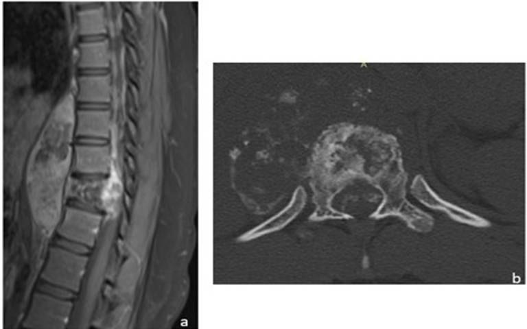
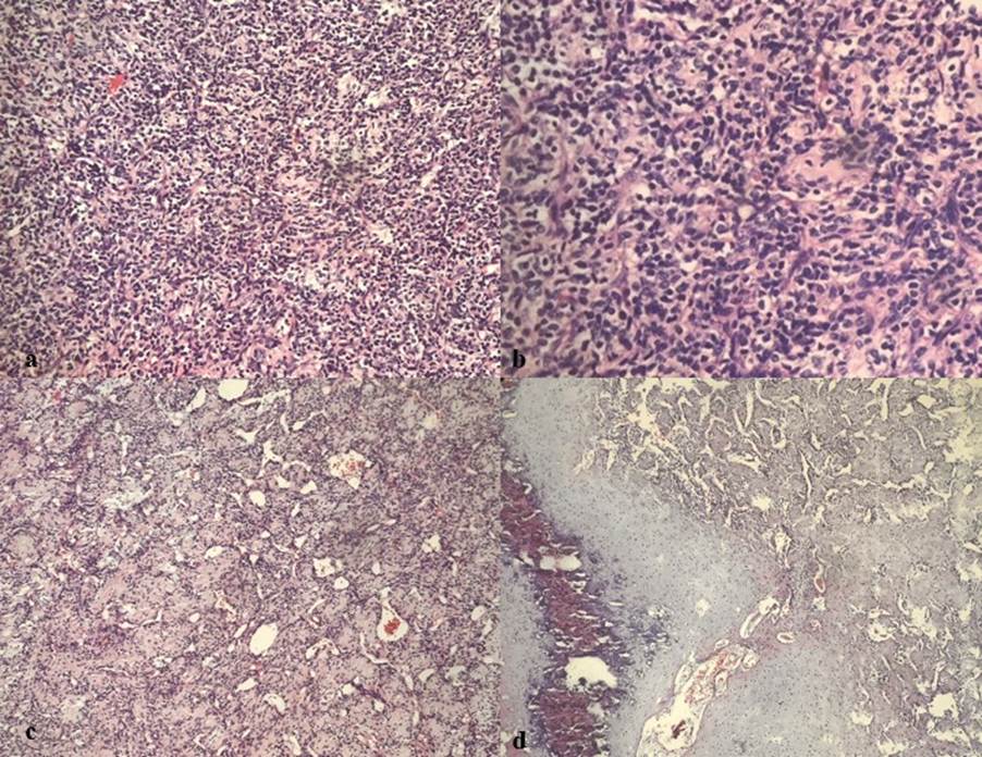
    <heading><span><bold>INTRODUCTION</bold></span></heading><p><span>Primary mesenchymal chondrosarcoma is a rare malignant neoplasm originating from bone or soft tissue<superscript>[<xref ref-type="link" rid="#ref-1">1</xref>]</superscript>. It is a subtype of chondrosarcoma which constitutes 3<superscript>rd </superscript>most common bone tumor and 10% of all chondrosarcomas<superscript>[<xref ref-type="link" rid="#ref-2">2</xref>]</superscript>. In the spine, thoracic vertebra is commonly involved and are extradural. Histologically these tumors are characterized by small round to ovoid malignant tumor cells mixed with foci of hyaline cartilage differentiation. Young adults have highest incidence with late local recurrence and metastasis<superscript>[<xref ref-type="link" rid="#ref-3">3</xref>]</superscript>. surgical resection is then treatment of choice. We present a case of mesenchymal chondrosarcoma in the thoracic vertebra which was simulating Pott’s spine clinically.</span></p><heading><span><bold>CASE REPORT</bold></span></heading><p><span>A 16 years-old female presented to neurology with chief complaints of back pain, weakness of lower limbs started as paraesthesia in both lower limbs since 15 days and difficulty in walking since 10 days. There is no history of fever, trauma, diabetes, hypertension or any exposure to Tuberculosis.</span></p><p><span>On examination patient is moderately built and nourished. No evidence of fever, pallor, icterus, cyanosis, clubbing, pedal edema or generalized lymphadenopathy. Tone of both lower limbs was increased (hypertonia). Meningeal signs were absent. Physical examination revealed both lower limb weakness (3/5). Hematological investigations were with in normal limits.</span></p><p><span>Chest X-ray PA view showed soft tissue density in right paravertebral region from D7-D10 vertebral body levels. CT spine dorsal plain showed collapse of D11 vertebral body with erosion. Adjacent pre and paravertebral soft tissue density with internal calcifications measuring 11.8X5.6X6.9cm was seen extending from D8 to D12 vertebral body levels. Posteriorly the soft tissue component with calcific foci measuring approximately 2.3X6.8mm is extending from D10-D11 levels in anterior epidural space, causing narrowing of the spinal canal at that level. Magnetic resonance imaging (MRI) of dorsal spine plain and contrast showed altered signal intensity lesion, which is predominantly hypo intense with few hyper intense areas in T1W1 and heterogeneously hyper intense inT2 with few hyperintensities showing heterogenous enhancement after contrast administration was seen involving D11 <xref ref-type="link" rid="#figure-1">[Fig. 1]</xref>. There is extension of posterior epidural soft tissue component into right neural foramina at D11 and D12 level. Above findings were highly suggestive of infective spondylitis likely tubercular etiology. </span></p><figure><graphic src="https://schoproductionportal.s3.ap-south-1.amazonaws.com/data/PIMR/239/1775474297112.jpeg"/><figcaption><span><bold>Fig. 1: a) T1 post contrast axial image showing heterogeneously enhancing altered signal intensity in D11 vertebral body (long arrow) with significant pre, right paravertebral (short arrow) and epidural (asterix) component. b) Non contrast CT axial images (soft tissue and bone window), sagittal reconstruction (bone window) are showing lytic and sclerotic destruction of D11 vertebra body (blue arrow) with pre, paravertebral, epidural soft tissue component with calcific foci (white arrow) </bold></span></figcaption></figure><p> </p><p><span>Surgical excision of tumor at D9, D10, D12 and L1 was done and was sent for histopathological examination. We received multiple grey-brown soft tissue bits together measuring 4.5X4X1.5cm. Microscopically, lesion was composed of round to spindle shaped cells having hyperchromatic nuclei and with scant to moderate amount of cytoplasm. Few foci showed heman- giopericytoma like vasculature. Islands of hyaline cartilage and foci of calcifications were also noted. With the above histological features, diagnosis of mesenchymal chondrosarcoma was considered <xref ref-type="link" rid="#figure-2">[Fig. 2]</xref>. </span></p><figure><graphic src="https://schoproductionportal.s3.ap-south-1.amazonaws.com/data/PIMR/239/1775474297188.jpeg"/><figcaption><span><bold>Fig. 2: a) Section showing lesion with small round to spindle shaped cells having hyperchromatic nuclei and with scant to moderate amount of cytoplasm (H&amp;E, X100). b) Section showing lesion with round to spindle shaped cells having hyperchromatic nuclei (H&amp;E, X400). c) Section showing hemangiopericytoma like vasculature and spindle shaped tumor cells (H&amp;E, X100). d) Section showing hemangiopericytoma like areas with islands of cartilage and foci of calcification (H&amp;E, 100)</bold></span></figcaption></figure><p> </p><heading><span><bold>DISCUSSION</bold></span></heading><p><span>Mesenchymal chondrosarcoma is a rare variant of chondrosarcoma accounting for 0.2% to 0.7% of malignant bone tumors<superscript>[<xref ref-type="link" rid="#ref-4">4</xref>]</superscript>. Mesenchymal chondrosarcoma was first reported by Lightenstein in 1959<superscript>[<xref ref-type="link" rid="#ref-5">5</xref>]</superscript>. These tumors originate probably from immature chondroblasts which will differentiate into mature cartilage, often with enchondral ossification<superscript>[<xref ref-type="link" rid="#ref-6">6</xref>]</superscript>.</span></p><p><span>Majority of tumors occur in second and third decade of life and does not show sex predilection. Maxilla and mandible are most commonly involved, but can also occur in pelvis, femur, ribs and spine<superscript>[<xref ref-type="link" rid="#ref-1">1</xref>]</superscript>. Intrathecal mesenchymal chondrsarcomas of spinal cord most commonly affects upper lumbar and lower thoracic spine. Approximately 1/3<superscript>rd</superscript> of these tumors are found in extraskeletal soft tissues, most commonly involving CNS and meninges. They can also involve soft tissue of the head and neck, extremities and visceral organs like female genitalia, pancreas, kidney, chest wall and retroperitoneal space<superscript>[<xref ref-type="link" rid="#ref-7">7</xref>-<xref ref-type="link" rid="#ref-9">9</xref>]</superscript>.</span></p><p><span>Most common genetic alteration observed in mesenchymal chondrosarcoma is the fusion of HEY1 and NCOA2 genes on chromosome 8(q13;q21). This fusion was reported by Wang et al in 2012 <superscript>[<xref ref-type="link" rid="#ref-10">10</xref>]</superscript> and is now used as molecular diagnostic marker for diagnosing mesenchymal chondrosarcomas which lack typical histological features<superscript>[<xref ref-type="link" rid="#ref-11">11</xref>]</superscript>.</span></p><p><span>Most of the patients with mesenchymal chondrosarcomas present with numbness and radicular pain due to compression of the lesion. Diagnosis of this condition is delayed due to non-specific clinical manifestation. Although imaging can help in diagnosis, it is difficult to distinguish this tumor from other conditions. On X-ray, mesenchymal chondrosarcomas present as osteolytic lesion with well-defined or indistinct cell borders or as soft tissue mass. On MRI, most of these tumors are isotense on T1W1 and T2W1 with well-defined borders and tend to be lobulated. In our case, lesion is hypotense with hyperintense areas in T1W1 and heterogeneously hyperintense on T2 suggesting infective etiology with possibility of Pott’s spine.</span></p><p><span>Histologically mesenchymal chondrosarcoma is a biphasic tumor composed of undifferentiated small round to spindle cells with hemangiopericytoma like vascular pattern and islands of mature appearing hyaline cartilage. On Immunohistochemistry (IHC), small round cells express CD99, NKX2.2 and cartilaginous component is positive for S-100. SOX-9 is positive in both cartilaginous component and small round cells. This tumor should be differentiated from other small round cell neoplasm like Ewing’s sarcoma, lymphoma, rhabdomyosarcoma and synovial sarcoma. </span></p><p><span>Ewing’s sarcoma lacks cartilaginous component and hemangiopericytoma like vasculature. On IHC, tumor cells express CD 99 and NKX2.2 but negative for SOX-9 and S-100. Lymphomas lack cartilaginous component. Tumor cells are positive for CD45 and are negative for S-100, NKX2.2, SOX-9. In Rhabdomyosarcoma, myogenin and Myo-D1 are expressed by tumor cells and are negative for CD 99, NKX2.2, SOX-9 and S-100. Synovial sarcoams does not have cartilagenous component and are positive for EMA, and TLE-1 but negative for S-100 and SOX-9. STAT6, and CD34 are expressed in solitary fibrous tumor but not in mesenchymal chondrosarcoma</span></p><p><span>Mesenchymal chondrosarcoma has high recurrence rate and metastasis Complete tumor resection along with stable spinal fixation is the treatment of choice for the tumor involving spine. Post-operative adjuvant radiotherapy can reduce the rate of local recurrence<superscript>[<xref ref-type="link" rid="#ref-12">12</xref>]</superscript>.</span></p><heading><span><bold>CONCLUSION</bold></span></heading><p><span>Mesenchymal chondrosarcoma is a rare malignant mesenchymal tumor located in bone, intracranial sites or soft tissue. As the imaging findings are not specific for mesenchymal chondrosarcoma, histopathology with additional immunohistochemistry is crucial for diagnosis. Due to high rate of recurrence and metastasis of these tumors, early diagnosis and long-term surveillance is recommended.</span></p><heading><span><bold>DISCLOSURE</bold></span></heading><p><span><bold>Source(s) of support:</bold> Nil.</span></p><p><span><bold>Conflicting Interest:</bold> Nil.</span></p>
  </body>
  <back>
    <ref-list>
      <title>References</title>
      
        
          <ref id="ref-1">
            <element-citation publication-type="journal">
              
                <person-group person-group-type="author">
                  <name>
                    Lin M, Zhou H, Zhang X, Hu Y, Guo T, Jitao SW
                  </name>
                </person-group>
              
              
                <article-title>Primary mesenchymal chondrosarcoma of the adult lumbar spine: a case report and review of the literature</article-title>
              
              
                <source>Translational Cancer Research</source>
              
              
                <year>2022</year>
              
              
                <volume>11</volume>
              
              
                <issue>9</issue>
              
              
                <uri>https://doi.org/10.21037/tcr-22-122</uri>
              
            </element-citation>
          </ref>


          <ref id="ref-2">
            <element-citation publication-type="journal">
              
                <person-group person-group-type="author">
                  <name>
                    Dehneh Y, Aldabbas M, Elfarissi MA, Khoulali M, Oulali N, Moufid F
                  </name>
                </person-group>
              
              
                <article-title>Spinal mesenchymal chondrosarcoma: A case report of a rare malignant tumor</article-title>
              
              
                <source>Surgical Neurology International</source>
              
              
                <year>2023</year>
              
              
                <volume>14</volume>


                <uri>https://doi.org/10.25259/sni_206_2023</uri>
              
            </element-citation>
          </ref>


          <ref id="ref-3">
            <element-citation publication-type="journal">
              
                <person-group person-group-type="author">
                  <name>
                    Zibis AH, Wade Shrader M, Segal LS
                  </name>
                </person-group>
              
              
                <article-title>Case Report: Mesenchymal Chondrosarcoma of the Lumbar Spine in a Child</article-title>
              
              
                <source>Clinical Orthopaedics &amp;amp; Related Research</source>
              
              
                <year>2010</year>
              
              
                <volume>468</volume>
              
              
                <issue>8</issue>
              
              
                <uri>https://doi.org/10.1007/s11999-010-1297-5</uri>
              
            </element-citation>
          </ref>


          <ref id="ref-4">
            <element-citation publication-type="journal">
              
                <person-group person-group-type="author">
                  <name>
                    Chen S, Wang Y, Su G, Shen B, Lin D
                  </name>
                </person-group>
              
              
                <article-title>Primary intraspinal dumbbell-shaped mesenchymal chondrosarcoma with massive calcifications: a case report and review of the literature</article-title>
              
              
                <source>World Journal of Surgical Oncology</source>
              
              
                <year>2016</year>
              
              
                <volume>14</volume>
              
              
                <issue>1</issue>
              
              
                <uri>https://doi.org/10.1186/s12957-016-0963-9</uri>
              
            </element-citation>
          </ref>


          <ref id="ref-5">
            <element-citation publication-type="journal">
              
                <person-group person-group-type="author">
                  <name>
                    Lightenstein L, Bernstein D
                  </name>
                </person-group>
              
              
                <article-title>Unusual benign and malignant chondroid tumors of bone.A survey of some mesenchymal cartilage tumors and malignant chondroblastic tumors, including a few multicentric ones, as well as many atypical benign chondroblastomas and chondromyxoid fibromas</article-title>
              
              
                <source>Cancer</source>
              
              
                <year>1959</year>
              
              
                <volume>12</volume>
              
              
                <issue>6</issue>
              
              
                <uri>https://doi.org/10.1002/1097-0142(195911/12)12:6&lt;1142::aid-cncr2820120610&gt;3.0.co;2-d</uri>
              
            </element-citation>
          </ref>


          <ref id="ref-6">
            <element-citation publication-type="journal">
              
                <person-group person-group-type="author">
                  <name>
                    Dudzisz-Sledz M, Kondracka M, Rudzinsk M, Zajac AE, Firlej W, Sulejczak D, &lt;I&gt;et al&lt;/I&gt;
                  </name>
                </person-group>
              
              
                <article-title>Mesenchymal Chondrosarcoma from Diagnosis to Clinical Trials</article-title>
              
              
                <source>Cancers</source>
              
              
                <year>2023</year>
              
              
                <volume>15</volume>
              
              
                <issue>18</issue>
              
              
                <uri>https://doi.org/10.3390/cancers15184581</uri>
              
            </element-citation>
          </ref>


          <ref id="ref-7">
            <element-citation publication-type="journal">
              
                <person-group person-group-type="author">
                  <name>
                    Lee Y, Choi S, Kim HS
                  </name>
                </person-group>
              
              
                <article-title>Extraskeletal Mesenchymal Chondrosarcoma of the Uterus</article-title>
              
              
                <source>Diagnostics</source>
              
              
                <year>2022</year>
              
              
                <volume>12</volume>
              
              
                <issue>3</issue>
              
              
                <uri>https://doi.org/10.3390/diagnostics12030643</uri>
              
            </element-citation>
          </ref>


          <ref id="ref-8">
            <element-citation publication-type="journal">
              
                <person-group person-group-type="author">
                  <name>
                    Esfahani MM, Mirazami SMA, Azadbakht J, Dashti F
                  </name>
                </person-group>
              
              
                <article-title>Retroperitoneal mesenchymal chondrosarcoma with metastasis to iliac vein: A rare case report and review of the literature</article-title>
              
              
                <source>Clinical Case Reports</source>
              
              
                <year>2022</year>
              
              
                <volume>10</volume>


                <uri>https://doi.org/10.22541/au.165884402.23565890/v1</uri>
              
            </element-citation>
          </ref>


          <ref id="ref-9">
            <element-citation publication-type="journal">
              
                <person-group person-group-type="author">
                  <name>
                    Goldenberg M, Ramgopal AA, Salgoda CM, Reyes-Mugica M, Malek MM, Tersak JM
                  </name>
                </person-group>
              
              
                <article-title>Mesenchymal chondrosarcoma of the chest wall in an adolescent patient: A case report and brief review of the literature</article-title>
              
              
                <source>Cancer Reports</source>
              
              
                <year>2022</year>
              
              
                <volume>5</volume>
              
              
                <issue>1</issue>
              
              
                <uri>https://doi.org/10.1002/cnr2.1453</uri>
              
            </element-citation>
          </ref>


          <ref id="ref-10">
            <element-citation publication-type="journal">
              
                <person-group person-group-type="author">
                  <name>
                    Wang L, Motoi T, Khanin R, Olshen A, Mertens F, Bridge J, &lt;I&gt;et al&lt;/I&gt;
                  </name>
                </person-group>
              
              
                <article-title>Identification of a novel, recurrent &lt;i&gt;HEY1‐NCOA2&lt;/i&gt; fusion in mesenchymal chondrosarcoma based on a genome‐wide screen of exon‐level expression data</article-title>
              
              
                <source>Genes, Chromosomes and Cancer</source>
              
              
                <year>2012</year>
              
              
                <volume>51</volume>
              
              
                <issue>2</issue>
              
              
                <uri>https://doi.org/10.1002/gcc.20937</uri>
              
            </element-citation>
          </ref>


          <ref id="ref-11">
            <element-citation publication-type="journal">
              
                <person-group person-group-type="author">
                  <name>
                    Moriya K, Katayama S, Onuma M, Rikiishi T, Hosaka M, Watanabe B, &lt;I&gt;et al&lt;/I&gt;
                  </name>
                </person-group>
              
              
                <article-title>Mesenchymal chondrosarcoma diagnosed on &lt;scp&gt;FISH&lt;/scp&gt; for &lt;i&gt;&lt;scp&gt;HEY1‐NCOA&lt;/scp&gt;&lt;/i&gt;&lt;i&gt;2&lt;/i&gt; fusion gene</article-title>
              
              
                <source>Pediatrics International</source>
              
              
                <year>2014</year>
              
              
                <volume>56</volume>
              
              
                <issue>5</issue>
              
              
                <uri>https://doi.org/10.1111/ped.12407</uri>
              
            </element-citation>
          </ref>


          <ref id="ref-12">
            <element-citation publication-type="journal">
              
                <person-group person-group-type="author">
                  <name>
                    Kawaguchi S, Weiss I, Lin PP, Huh WW, Lewis VO
                  </name>
                </person-group>
              
              
                <article-title>Radiation Therapy Is Associated With Fewer Recurrences in Mesenchymal Chondrosarcoma</article-title>
              
              
                <source>Clinical Orthopaedics &amp;amp; Related Research</source>
              
              
                <year>2014</year>
              
              
                <volume>472</volume>
              
              
                <issue>3</issue>
              
              
                <uri>https://doi.org/10.1007/s11999-013-3064-x</uri>
              
            </element-citation>
          </ref>
        
      
    </ref-list>
  </back>
</article>