SciresolSciresolhttps://jmsh.ac.in/Journal of Medical Sciences and Health10.47799/pimr.1402.26.88ORIGINAL ARTICLE<p>Clinicopathological Spectrum of Non-Neoplastic Lesions of Skin Punch Biopsies: A Retrospective Study</p>303202620261421Abstract
<p>Non-neoplastic skin lesions are more prevalent than neoplastic lesions. Clinically, many skin lesions exhibit overlapping morphological features, making correct diagnosis difficult without histopathological evaluation. A pattern-based approach to evaluation of skin punch biopsies facilitates categorization and diagnosis.<bold> Aim and Objectives:</bold> To analyze the clinical presentation, histopathological spectrum of non-neoplastic skin lesions and classify them based on tissue reaction patterns.<bold> Materials and Methods: </bold>This retrospective study was conducted over one year (January–December 2025) in the Department of Pathology at a tertiary care center. Relevant clinical data were retrieved, histopathological slides were examined, diagnoses were established and categorized according to tissue reaction patterns. Findings were compiled and analyzed in Microsoft Excel.<bold> Results: </bold>A total of 88 skin punch biopsies were studied: 45 females (51.1%) and 43 males (48.9%). The most frequently affected age group was 31-40 years (26.1%) followed by 41-50 years (21.6%) Common presentations included hyperpigmented plaques/patches (33%), erythematous lesions (23.9%), hypopigmented lesions (12.5%) and itchy plaques (13.6%). Among major tissue reaction patterns our study found Lichenoid pattern (19.3%) to be highest in number within which lichen planus (4) was most frequent. Followed by psoriasiform pattern (18.2%) within which prurigo nodularis (6) was most frequent then granulomatous pattern (9.1%). 11.4% cases were categorized under nonspecific lesions as few of them had non-specific findings and few others were inadequately sampled.<bold> Conclusion: </bold>Histopathological tissue reaction pattern analysis along with clinical features helps in categorization which streamlines the diagnosis of non-neoplastic skin lesions in punch biopsies.</p>
KeywordsSkinPunch biopsyLichenoidGranulomatousLeprosyPsoriasiformPathangeDeepak1 Assistant Professor, Malla Reddy Institute of Medical Sciences Malla Reddy Vishwavidyapeeth (Deemed to be University) Hyderabad, Telangana India Professor, Malla Reddy Institute of Medical Sciences Malla Reddy Vishwavidyapeeth (Deemed to be University) Hyderabad, Telangana India Professor and HOD, Malla Reddy Institute of Medical Sciences Malla Reddy Vishwavidyapeeth (Deemed to be University) Hyderabad, Telangana IndiaChennupatiVenkata Sri Laxmi1 Assistant Professor, Malla Reddy Institute of Medical Sciences Malla Reddy Vishwavidyapeeth (Deemed to be University) Hyderabad, Telangana India Professor, Malla Reddy Institute of Medical Sciences Malla Reddy Vishwavidyapeeth (Deemed to be University) Hyderabad, Telangana India Professor and HOD, Malla Reddy Institute of Medical Sciences Malla Reddy Vishwavidyapeeth (Deemed to be University) Hyderabad, Telangana IndiaBalinaLakshmi Nirmala1 Assistant Professor, Malla Reddy Institute of Medical Sciences Malla Reddy Vishwavidyapeeth (Deemed to be University) Hyderabad, Telangana India Professor, Malla Reddy Institute of Medical Sciences Malla Reddy Vishwavidyapeeth (Deemed to be University) Hyderabad, Telangana India Professor and HOD, Malla Reddy Institute of Medical Sciences Malla Reddy Vishwavidyapeeth (Deemed to be University) Hyderabad, Telangana IndiaKasturiSumalatha2 Assistant Professor, Malla Reddy Institute of Medical Sciences Malla Reddy Vishwavidyapeeth (Deemed to be University) Hyderabad, Telangana India Professor, Malla Reddy Institute of Medical Sciences Malla Reddy Vishwavidyapeeth (Deemed to be University) Hyderabad, Telangana India Professor and HOD, Malla Reddy Institute of Medical Sciences Malla Reddy Vishwavidyapeeth (Deemed to be University) Hyderabad, Telangana IndiaShastrySrikanth3 Assistant Professor, Malla Reddy Institute of Medical Sciences Malla Reddy Vishwavidyapeeth (Deemed to be University) Hyderabad, Telangana India Professor, Malla Reddy Institute of Medical Sciences Malla Reddy Vishwavidyapeeth (Deemed to be University) Hyderabad, Telangana India Professor and HOD, Malla Reddy Institute of Medical Sciences Malla Reddy Vishwavidyapeeth (Deemed to be University) Hyderabad, Telangana IndiaINTRODUCTION
Skin disorders represent one of the most common health concerns worldwide. Non-neoplastic lesions of the skin are considerably more prevalent than neoplastic lesions[1, 2]. Research conducted across the Asian subcontinent consistently indicates a significant burden of skin diseases, with a wide and diverse spectrum[3]. Clinically, many of these lesions exhibit overlapping morphological features, making correct diagnosis challenging without histopathological evaluation[2, 4]. Histopathological examination plays a crutial role in differentiating and confirming these lesions[1-3].
A pattern-based approach to the histopathological assessment of skin punch biopsies facilitates categorization and diagnosis. Establishing correct diagnosis is crucial as it guides clinicians in determining prognosis, selecting appropriate therapeutic interventions, and planning the subsequent management of these dermatological conditions[1].
The present study aimed to analyse the clinical presentation and histopathological spectrum of non-neoplastic skin lesions diagnosed on punch biopsy and to categorise these lesions according to their predominant histopathological tissue reaction patterns.
MATERIALS AND METHODS
Study Design and Participants: This retrospective observational study was conducted over a period of one year from January 1st 2025 to December 31st 2025 in the Department of Pathology of a tertiary care centre hospital. The study included skin punch biopsy specimens received in the histopathology laboratory during the study period for evaluation of clinically suspected non-neoplastic dermatoses. Biopsies submitted with a clinical differential diagnosis of non-neoplastic dermatoses were included. Each biopsy represented a separate patient.
Inclusion and Exclusion Criteria: All skin punch biopsies submitted to the histopathology laboratory with a clinical differential diagnosis of non-neoplastic dermatoses were included in the study. All eligible cases received during the study period were included consecutively. All benign and malignant neoplastic skin lesions were excluded from the study.
Data Collection and Variables: Relevant clinical and demographic data were retrieved from the medical records including age, sex, clinical presentation. Histopathological variables included the predominant tissue reaction pattern and the specific histopathological diagnosis. The data were obtained from both Medical Records Department and histopathology records and were subsequently compiled for analysis.
Histopathological examination:All biopsies which were obtained under local anaesthesia using aseptic conditions were fixed in 10% neutral buffered formalin and subsequently processed in the histopathology laboratory. Tissue samples underwent routine paraffin embedding followed by microtome sectioning and staining with Haematoxylin and Eosin (H&E). Special stains, including Fite-Faraco and acid-fast bacilli (AFB) stains were employed whenever indicated. Histopathological slides were examined and diagnoses were established based on microscopic findings and categorised based on tissue reaction patterns.
Tissue Reaction Patterns: Histopathological diagnoses were categorized according to the predominant tissue reaction pattern identified on microscopic examination as described in Weedon’s skin pathology[1]. The major patterns included lichenoid, granulomatous, psoriasiform, vasculopathic, spongiotic and vesiculobullous patterns. Infectious dermatoses and other histopathological categories that did not fall under the major tissue reaction patterns were classified separately. In cases where more than one tissue reaction patterns were observed were categorized based on the predominant pattern, determined by relative extent and prominence of characteristic histopathological features. Cases showing non-specific or insufficient histopathological findings were categorized separately where a definitive tissue reaction pattern could not be assigned.
Statistical Analysis: Data were entered and analysed using Microsoft Excel. Categorical variables were summarized as frequencies and percentages.
Ethical Considerations: The study was conducted after obtaining approval from the Institutional Ethics Committee (approval number: FAC/2026/173, dated 20 February 2026). As this was a retrospective study based on existing records and archived histopathological material, the requirement for individual informed consent was waived in accordance with institutional policy.
RESULTS
A total of 88 skin punch biopsy specimens were analysed. Of these, 45 (51.1%) were from female patients and 43 (48.9%) were from male patients, giving a male-to-female ratio of 0.95:1. The most frequently affected age group was between 31–40 years (23 cases, 26.1%) followed by 41-50 age group (19 cases,21.6%) as shown in [Table. 1].
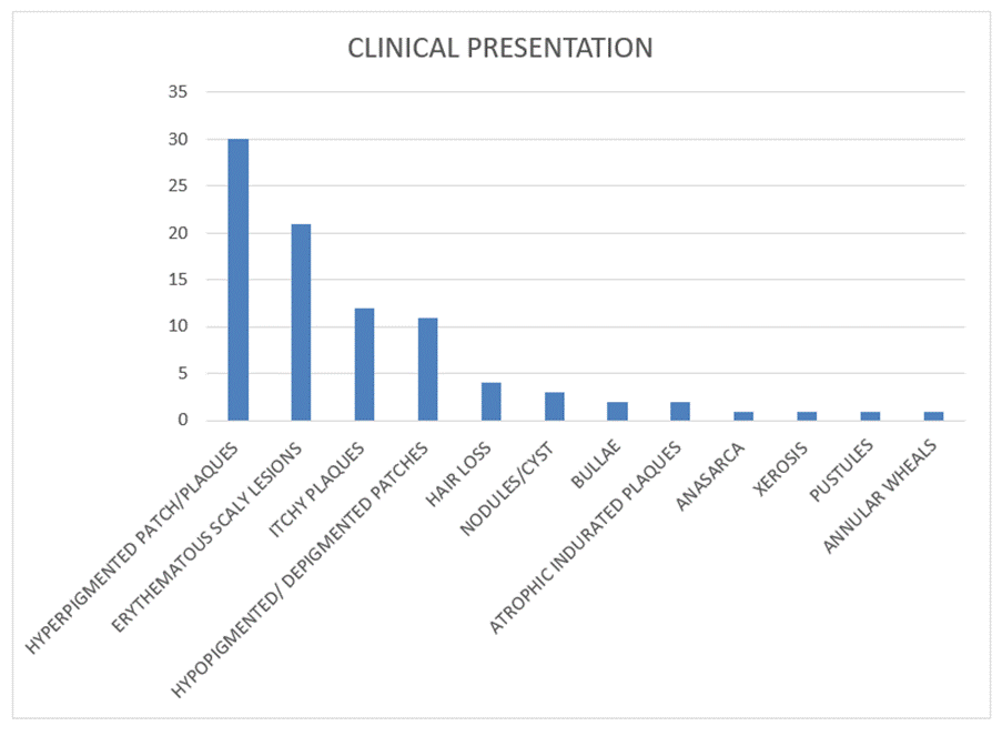
Based on clinical history, cases were categorized according to their morphological appearance. Most common were hyperpigmented plaques or patches (29 cases, 33%), hypopigmented or depigmented lesions (11 cases, 12.5%), erythematous lesions (21 cases, 23.9%), itchy plaques (12 cases, 13.6%). The remaining less frequent presentations are mentioned in [Fig. 1].
Based on histopathological examination, out of 88 cases, 78 (88.6%) were classified according to the predominant tissue reaction pattern. The remaining 10 cases (11.4%) were put under nonspecific category because of nonspecific histopathological findings (8) and biopsy sent was inadequate to comment under microscopy(2) ([Table. 2] and [Table. 3]).
Among all the major tissue reaction patterns, 17 cases were showing Lichenoid pattern which was found to be highest in number followed by 16 cases showing psoriasiform pattern, granulomatous pattern was observed in 8 cases. 3 cases were showing vasculopathic pattern, 1 case each showing spongiotic and vesiculobullous patterns.
Lichenoid Pattern group (17 cases, 19.3%):This tissue reaction pattern included all cases which showed microscopic features of epidermal basal cell damage, Interface dermatitis, band like infiltrate of inflammatory cells in superficial dermis, pigment incontinence and Civatte bodies. Lichen planus (4 cases) was most common followed by Lichen planopilaris (3 cases) which histologically showed a Lichenoid reaction involving upper follicular epithelium (infundibulum and isthmus) with dense perifollicular infiltrate (lymphocytes and macrophages) and 3 cases were of Erythema dyschromicum perstans which showed melanin incontinence in dermis. Hypertrophic lichen planus and discoid lupus erythematosus each constituted 2 cases, while Fixed drug eruption, lichen sclerosus et atrophicus and Photosensitive lichenoid eruption were noted in 1 case each.
Age group(years)
No. of. cases (%)
0-10
02(2.2)
11-20
17(19.3)
21-30
16(18.2)
31-40
23(26.1)
41-50
19(21.6)
51-60
08(9.1)
61-70
01(1.1)
71-80
02(2.2)
Total
88
Table 1: Number of cases according to age groups
Fig. 1: Representation of clinical presentation of all skin lesions
Psoriasiform Pattern (16 cases, 18.2%) showed epidermal hyperplasia, rete ridge elongation, regular acanthosis. Within this group prurigo nodularis (6 cases) showed irregular epidermal hyperplasia, marked acanthosis. 2 out of 6 cases of prurigo nodularis showed focal pseudoepitheliomatous hyperplasia, they were categorized under psoriasiform pattern because this was predominant pattern and pseudoepitheliomatous hyperplasia was considered as secondary or minor tissue pattern. Pityriasis rosea (4 cases) which showed psoriasiform hyperplasia, spongiosis, dermal lymphocytic infiltrate followed by psoriasis (2 cases) which showed elongation of rete ridges, collection of neutrophils in stratum corneum (Munro’s abscess). Lichen simplex chronicus (2 cases) showed features of psoriasiform hyperplasia, vertical streaking of collagen in papillary dermis. 1 case each of exfoliative dermatitis and spongiotic dermatosis were observed. Spongiotic dermatosis showed predominant tissue pattern of psoriasiform epidermal hyperplasia with focal spongiosis hence it was categorised under psoriasiform and not under spongiotic pattern.
Papular urticaria (1), small vessel vasculitis (1), vasculitis with subcorneal pustules (1)
TOTAL
46(52.2)
Table 2: Frequency of major tissue reaction patterns with specific diseases
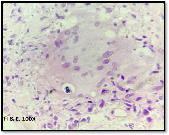
Granulomatous Pattern (8 cases, 9.1%): Tuberculoid leprosy (1 case) showed well-formed epithelioid granulomas involving dermis, around adnexa and nerves. Borderline tuberculoid leprosy (3 cases) showed dermal peri adnexal lymphocytic and epithelioid collections along with nerve bundle thickening. Other granulomatous dermatoses included tattoo granuloma (1 case) which showed black pigment laden macrophages, granulomatous cheilitis (1 case) which showed non caseating granulomas in dermis and granulomatous dermatitis of sarcoidal type (2 cases) which showed features of diffuse epithelioid granulomas, a few Langhans giant cells with intracytoplasmic concretions i.e. Schaumann bodies. These cases were negative for PAS, AFB and Fite -Faraco stains.
Other major histopathological patterns [Table. 2] such as vasculopathic pattern (3.4%) which showed endothelial damage, perivascular inflammatory infiltrate, fibrinoid necrosis, leukocytoclasia. 1 case each of papular urticaria, small vessel vasculitis and vasculitis with subcorneal pustules were categorized under this pattern. Spongiotic pattern (1.1%) with spongiosis, prominent intercellular spaces were observed in 1 case of subacute eczema. In vesiculobullous pattern (1.1%) a case of bullous pemphigoid with subepidermal bulla was observed.
Table 3: Frequency of other histopathological categories and associated diagnoses
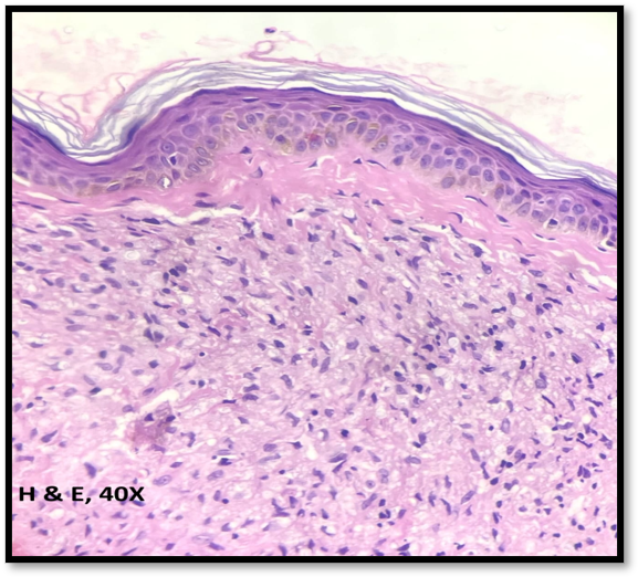
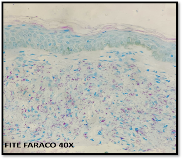
Under Infectious dermatoses [Table. 3], a total of 11 cases (12.5%) were identified which included 2 cases of verruca vulgaris with papillomatosis and inward bending of rete ridges, 1 case of fungal infection showed few yeast forms and fungal hyphae, 5 cases of Lepromatous leprosy histologically showed a Grenz zone, diffuse infiltration of histiocytes and globi formation. On Fite-Faraco staining, out of 05 cases of lepromatous leprosy, 03 cases showed bacillary index of 5, remaining 02 cases showed bacillary index of 6. 1 case of Borderline lepromatous leprosy showed foamy macrophages in the dermis, 2 cases of Indeterminate leprosy showed perivascular and peri adnexal lymphocytic infiltration.
Morphea (5 Cases) showed thickened homogenised densely packed collagen bundles, and these were categorised under collagen disorders. Under diseases of cutaneous appendages (5,5.7%), Folliculitis (1 case) showed perifollicular neutrophilic inflammation, Folliculitis decalvans (2 cases) showed neutrophilic folliculitis with follicular destruction and Pseudopelade of Brocq (2 cases) showed follicular loss with fibrosis and minimal inflammation was observed. Under panniculitis, Septal panniculitis (1 case) showed inflammation involving septa of subcutaneous fat and Erythema nodosum leprosum (1 case) showed predominant subcutaneous neutrophilic inflammation in septa and lobules, leukocytoclasia in the background of lepromatous inflammation. ENL was categorized it into panniculitis type of lesion because predominant histopathological findings were those of panniculitis. The other less frequent entities were documented as shown in [Table. 3].
Out of 10 remaining cases which could not be subtyped in any of the above described tissue reaction patterns, 8 cases with clinical suspicion of Hansen’s disease(5cases), pityriasis rosea (1case) and lichen planus (2 cases) showed only mild perivascular and peri adnexal chronic inflammatory infiltrate. These findings were inadequate to establish a definitive diagnosis and hence categorized under nonspecific. 2 cases showed only epidermis, dermis and subcutis were not included in biopsy and hence were inadequate for interpretation.
DISCUSSION
In the present study, a total of 88 cases of non-neoplastic skin lesions were collected and analysed retrospectively. This sample size was comparable to those reported in previous studies; Singh et al. analysed 112 cases retrospectively over 2 years, while Veldurthy et al., Mehar et al., Chinnathambi et al. reported 97,112 and 82 cases, respectively over a period of one year[5-8].
Our study showed slight female preponderance. Interestingly, most other studies reported a male preponderance. For instance, Singh et al. observed 54.5% males and 45.5% females, Mittal et al. reported 60% males and 40% females and Chinnathambi et al. documented 56.09% males and 43.9% females[5, 8, 9]. As the present study was based on skin punch biopsies received at a tertiary care centre rather than a population-based survey, the observed sex distribution may reflect differences in referral and biopsy practices rather than the actual prevalence of non-neoplastic skin diseases in the general population.
The most commonly affected age group in our study was 31-40 years. This observation aligns with the findings of Reddy et al. and Gupta et al., who also reported the highest incidence in the fourth decade of life[10, 11]. In contrast, Veldurthy et al. and Younas et al. found the majority of cases in the 21–30 years age group[6, 12].
The most common clinical presentation in our study was hyperpigmented plaques or patches followed by erythematous lesions and hypopigmented or depigmented lesions. This is in contrast to study by Mittal et al who observed more hypopigmented lesions followed by hyperpigmented lesions in his study cohort[9].
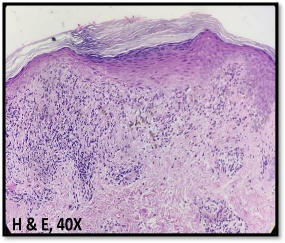
In the present study, the most frequent histopathological patterns encountered were the lichenoid tissue reaction pattern. This reflects the wide histopathological spectrum of inflammatory dermatoses encountered in routine dermatopathology practice. When compared with previous studies, certain variations were noted. Singh et al. reported non-specific dermatoses as the most common histopathological finding, followed by granulomatous lesions[5]. Veldurthy et al. observed lichenoid lesions as the most frequent category, followed by Hansen’s disease which correlates with spectrum and prevalence of leprosy in their study population[6]. Reddy et al. documented psoriasiform lesions as the predominant pattern, accounting for 42.5% of cases, followed by lichen planus, highlighting regional and demographic differences in the occurrence of specific dermatoses[10]. On the other hand, Mehar et al. found granulomatous lesions to be the most common pattern, followed by non-specific dermatoses supporting the observation that granulomatous reactions remain a significant diagnostic category in inflammatory skin pathology[7]. The distribution of lesion types shows variability across different populations and geographic regions. This variation may be due to environmental factors, genetic predisposition, socio-economic conditions and differences in the prevalence of infectious dermatoses such as Hansen’s disease. Typical example of Lichenoid tissue reaction is lichen planus shown in [Fig. 2]. Lichen planus was also the most common entity among lichenoid pattern of tissue reaction in studies by Reddy et al, Singh and Kanwar, Bhattacharya et al., Abdallat and Maaita, and Anbar et al[13-17].
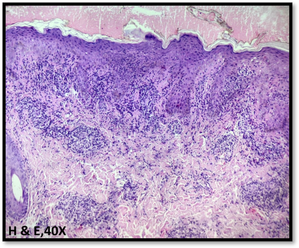
The most common entity with psoriasiform tissue reaction was Prurigo nodularis followed by Pityriasis rosea, psoriasis and lichen simplex chronicus whereas a study by Leelama JP et al showed most common entity was psoriasis followed by lichen simplex chronicus and prurigo nodularis[18]. This can be attributed to the above study included only psoriasiform patterned lesions. A study on erythematous plaques by Reddy et al showed psoriasis (42.5%) followed by lichen planus[10]. Mittal et al showed 2 cases psoriasis in their study of total100 cases[9]. A case of Pityriasis rosea as shown in [Fig. 3].
Fig. 2: Lichen Planus H&E40X: Epidermis showing hyperkeratosis, hypergranulosis, irregular acanthosis, basal cell vacuolation. Band like lymphocytic infiltrate at dermoepidermal junction, melanin incontinence noted
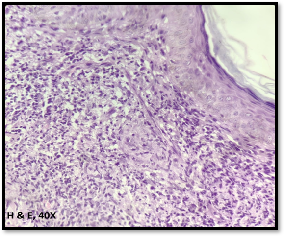
Fig. 4: Sarcoidosis H&E 40X: Dermis showing diffuse epithelioid granulomas, few Langhans giant cell surrounded by lymphocytes
Among granulomatous tissue reaction, borderline tuberculoid leprosy was more common than tuberculoid leprosy whereas Mittal et al. identified tuberculoid leprosy as the most frequent subtype followed by indeterminate and lepromatous leprosy. However lepromatous leprosy was included in granulomatous tissue reaction pattern in above study whereas it was included under infectious dermatosis in the current study[9].Veldurthy et al. reported 5 cases of tuberculoid leprosy and 3 cases of borderline tuberculous out of total 92 cases[6]. As other types of leprosy, borderline lepromatous, indeterminate and lepromatous leprosy showed no granulomas they were categorized under infectious type of lesions of skin in the present study. Other entities included in this tissue reaction were of granulomatous dermatitis of sarcoid type as seen in ([Fig. 4] and [Fig. 5].
Fig. 5: Sarcoidosis H&E 100X: One giant cell showing small basophilic intracytoplasmic concretions- Schaumann body
Fig. 6: Lepromatous Leprosy H&E 40X: Loss of rete ridges, Grenz zone in dermo epidermal junction. Dermis showing sheets of foamy histiocytes
Under infectious dermatosis, Indeterminate leprosy, borderline lepromatous leprosy and lepromatous leprosy were categorized. Our study corresponds with veldhurty et al which showed 5 cases of inderterminate, 2 cases borderline lepromatous leprosy and 5 cases of lepromatous leprosy out of total 92 cases in the study[6]. However, Singh et al showed only 1 case of borderline leprosy and 2 cases of lepromatous leprosy out of 112 cases in total, suggesting the regional differences in the spectrum of Hansen’s disease presentations[5]. Mehar et al considered all lepromatous lesions under granulo- matous which exaggerated their number to 48 cases in total out of 112[7]. A case of lepromatous leprosy [Fig. 6]. Fite-Faraco staining of lepromatous leprosy exhibiting a bacillary index of 6 as shown in [Fig. 7].
Fig. 7: Lepromatous Leprosy Fite-Faraco 40X: Highlights acid fast bacilli, Bacillary index 6
Although ENL is considered as infectious or granulomatous type, in our study we categorized it into panniculitis type of lesion because panniculitis was the predominant histopathological pattern. This is an example of why reaction pattern-based categorization based on predominant pattern is useful when there is more than one pattern in the biopsy.
Limitations: As it is a retrospective study, clinicopatho- logical correlation was dependent on completeness of clinical information documented in the records as pattern-based assessment fails when decoupled from clinical data. Some individual reaction pattern categories contained only a small number of cases limiting meaningful subgroup comparisons.
CONCLUSION
Histopathological examination of non-neoplastic skin punch biopsies demonstrates a broad spectrum of tissue reaction patterns, with considerable overlap among different dermatological disorders. A systematic pattern-based approach enables recognition of the predominant tissue response and facilitates a focused differential diagnosis. However correct diagnosis requires correlation of histopathological findings with clinical presentation. Thus, familiarity with tissue reaction patterns combined with effective communication between pathologist and dermatologist can facilitate in diagnosis and reduce diagnostic errors. As leprosy emerged as one of the frequent diagnoses it highlights leprosy as a public health problem at the national level, the disease continues to occur and remains relevant in routine dermatopatho- logical practice.
References
Calonje E, Bhogal BS
Histopathology of the Skin: General PrinciplesRook's Textbook of Dermatology, Ninth Edition2016https://doi.org/10.1002/9781118441213.rtd0003
Singh R, Bharathi K, Bhat R, Udayashankar C
The histopathological profile of non-neoplastic dermatological disorders with special reference to granulomatous lesions: study at a tertiary care centre in PondicherryThe Internet Journal of Pathology2012133https://doi.org/10.5580/2bf9
Veldurthy VS, Shanmugam C, Sudhir N, Sirisha O, Motupalli CP, Rao N, <I>et al</I>
Pathological study of non-neoplastic skin lesions by punch biopsyInternational Journal of Research in Medical Sciences201538https://doi.org/10.18203/2320-6012.ijrms20150313
Mehar R, Jain R, Kulkarni CV, Narang S, Mittal M, Patidar H
Histopathological study of dermatological lesions - A retrospective approachInternational Journal of Medical Science and Public Health201439https://doi.org/10.5455/ijmsph.2014.190620142
Sakthidasan Chinnathambi P, Burra A
Histopathological study of cutaneous non-neoplastic lesions in punch biopsiesIP Indian Journal of Clinical and Experimental Dermatology202172https://doi.org/10.18231/j.ijced.2021.025
Mittal A, Ahmad F, Bharadwaj K, Awasthi S, Dutta S
Histopathological study of non-neoplastic skin lesions-A retrospective approachIndian Journal of Pathology and Oncology201964https://doi.org/10.18231/j.ijpo.2019.108
Reddy R, Krishna N
Histopathological spectrum of non-infectious erythematous, papulo-squamous lesionsAsian Pacific Journal of Health Sciences201414Shttps://doi.org/10.21276/apjhs.2014.1.1s.6
Gupta I, Kaira V, Gupta K, Bothale KA, Mahore SD
Clinical profile of non neoplastic skin lesions: A prospective cross-sectional studyIP Indian Journal of Clinical and Experimental Dermatology201952https://doi.org/10.18231/j.ijced.2019.034
Younas M, Haque A
Spectrum of histopathological features in non-infectious erythematous and papulosquamous diseasesInternational Journal of Pathology200421https://jpathology.com/index.php/OJS/article/view/16
Reddy PKS, Sumathy TK, Shyamprasad AL, Shivaswamy KN, Suparna MY
Clinical, dermoscopic, and histopathological correlation of lichenoid dermatosesIndian Journal of Dermatopathology and Diagnostic Dermatology201962https://doi.org/10.4103/ijdpdd.ijdpdd_71_18
Singh OP, Kanwar AJ
Lichen planus in India: an appraisal of 441 casesInternational Journal of Dermatology19761510https://doi.org/10.1111/j.1365-4362.1976.tb00175.x
Bhattacharya M, Kaur I, Kumar B
Lichen Planus: A Clinical and Epidemiological StudyThe Journal of Dermatology2000279https://doi.org/10.1111/j.1346-8138.2000.tb02232.x
Anbar TE, Barakat M, Ghannam SF
A clinical and epidemiological study of lichen planus among Egyptians of AL-Minya provinceDermatology Online Journal2005112https://doi.org/10.5070/d30b91t45n
Leelamma JP, Babitha AM, Sankar S, Nandakumar G
Histopathological spectrum of Psoriasiform dermatitisJournal of Pathology of Nepal2016612https://doi.org/10.3126/jpn.v6i12.16265