<?xml version='1.0' encoding='UTF-8'?>

<!DOCTYPE article PUBLIC "-//NLM//DTD JATS (Z39.96) Journal Publishing DTD v1.1d1 20130915//EN" "JATS-journalpublishing1.dtd">
<article xmlns:xlink="http://www.w3.org/1999/xlink">
  <front>
    <journal-meta id="journal-meta-87cddb9ab7774ac9973b6a64b7cbc767">
      <journal-id journal-id-type="nlm-ta">Sciresol</journal-id>
      <journal-id journal-id-type="publisher-id">Sciresol</journal-id>
      <journal-id journal-id-type="journal_submission_guidelines">https://jmsh.ac.in/</journal-id>
      <journal-title-group>
        <journal-title>Journal of Medical Sciences and Health</journal-title>
      </journal-title-group>
      <issn publication-format="print"/>
    </journal-meta>
    <article-meta>
        
          
            <article-id pub-id-type="doi">10.47799/pimr.1402.26.89</article-id>
          
          
            <article-categories>
              <subj-group>
                <subject>CASE REPORT</subject>
              </subj-group>
            </article-categories>
            <title-group>
              <article-title>&lt;p&gt;Subcutaneous Fungal Granuloma Clinically Mimicking Malignancy: A Rare Case Report&lt;/p&gt;</article-title>
            </title-group>
          
          
            <pub-date date-type="pub">
              <day>30</day>
              <month>3</month>
              <year>2026</year>
            </pub-date>
            <permissions>
              <copyright-year>2026</copyright-year>
            </permissions>
          
          
            <volume>14</volume>
          
          
            <issue>2</issue>
          
          <fpage>1</fpage>

          <abstract>
            <title>Abstract</title>
            &lt;p&gt;&lt;bold&gt;Background: &lt;/bold&gt;&lt;span&gt;Subcutaneous fungal granulomas are rare deep fungal infections that often mimic neoplastic or inflammatory lesions, making diagnosis challenging, particularly in immunocompetent elderly individuals. Their nonspecific clinical presentation frequently results in delayed diagnosis and inappropriate initial management. &lt;/span&gt;&lt;bold&gt;Case Details: &lt;/bold&gt;&lt;span&gt;This is a 74-year-old female presented with a gradually progressive painful swelling over the right thigh for three months. Clinical examination suggested a soft tissue sarcoma, lipoma, abscess, and malignancy considered as differential diagnoses. MRI demonstrated hypertrophied edematous muscles, while FNAC was suggestive of a giant cell-containing lesion. Histopathological examination revealed necrotizing granulomatous inflammation with broad ribbon-like fungal hyphae, establishing the diagnosis of subcutaneous fungal granuloma. Following histopathological diagnosis, the patient was initially prescribed itraconazole 200 mg orally once daily for 3 days and on follow-up, there was clinical improvement in the swelling and associated pain, and the patient was reported to be clinically well without significant ongoing symptoms. &lt;/span&gt;&lt;bold&gt;Conclusion: &lt;/bold&gt;&lt;span&gt;Subcutaneous fungal granuloma should be considered in the differential diagnosis of chronic soft tissue swellings, even in immunocompetent elderly patients. Early clinicopathological correlation and histopathological evaluation are essential for timely diagnosis and appropriate management.&lt;/span&gt;&lt;/p&gt;
          </abstract>
          
          
            <kwd-group>
              <title>Keywords</title>
              
                <kwd>Subcutaneous fungal granuloma; Deep mycosis; Granulomatous inflammation</kwd>
              
            </kwd-group>


        <contrib-group>
          
            
              <contrib contrib-type="author">
                <name>
                  <surname>Japa</surname>
                  <given-names>Sai Krishna Reddy</given-names>
                </name>
                
                  <xref rid="aff-1" ref-type="aff">1</xref>
                
              </contrib>


              <aff id="aff-1">
                <institution> MBBS Prathima Institute of Medical Sciences </institution>
                <addr-line>Nagunur, Karimnagar-505417, Telangana India</addr-line>
              </aff>
            
              <aff id="aff-2">
                <institution> Final MBBS Part-II Student Prathima Institute of Medical Sciences </institution>
                <addr-line>Nagunur, Karimnagar-505417, Telangana India</addr-line>
              </aff>
            
              <aff id="aff-3">
                <institution> Professor, Department of Pathology Prathima Institute of Medical Sciences </institution>
                <addr-line>Nagunur, Karimnagar-505417, Telangana India</addr-line>
              </aff>
            
              <aff id="aff-4">
                <institution> Professor &amp; Head, Department of Pathology Prathima Institute of Medical Sciences </institution>
                <addr-line>Nagunur, Karimnagar-505417, Telangana India</addr-line>
              </aff>


              <contrib contrib-type="author">
                <name>
                  <surname>Varaganti</surname>
                  <given-names>Vamshi</given-names>
                </name>
                
                  <xref rid="aff-2" ref-type="aff">2</xref>
                
              </contrib>


              <aff id="aff-1">
                <institution> MBBS Prathima Institute of Medical Sciences </institution>
                <addr-line>Nagunur, Karimnagar-505417, Telangana India</addr-line>
              </aff>
            
              <aff id="aff-2">
                <institution> Final MBBS Part-II Student Prathima Institute of Medical Sciences </institution>
                <addr-line>Nagunur, Karimnagar-505417, Telangana India</addr-line>
              </aff>
            
              <aff id="aff-3">
                <institution> Professor, Department of Pathology Prathima Institute of Medical Sciences </institution>
                <addr-line>Nagunur, Karimnagar-505417, Telangana India</addr-line>
              </aff>
            
              <aff id="aff-4">
                <institution> Professor &amp; Head, Department of Pathology Prathima Institute of Medical Sciences </institution>
                <addr-line>Nagunur, Karimnagar-505417, Telangana India</addr-line>
              </aff>


              <contrib contrib-type="author">
                <name>
                  <surname>Sharadrutha</surname>
                  <given-names>A</given-names>
                </name>
                
                  <xref rid="aff-3" ref-type="aff">3</xref>
                
              </contrib>


              <aff id="aff-1">
                <institution> MBBS Prathima Institute of Medical Sciences </institution>
                <addr-line>Nagunur, Karimnagar-505417, Telangana India</addr-line>
              </aff>
            
              <aff id="aff-2">
                <institution> Final MBBS Part-II Student Prathima Institute of Medical Sciences </institution>
                <addr-line>Nagunur, Karimnagar-505417, Telangana India</addr-line>
              </aff>
            
              <aff id="aff-3">
                <institution> Professor, Department of Pathology Prathima Institute of Medical Sciences </institution>
                <addr-line>Nagunur, Karimnagar-505417, Telangana India</addr-line>
              </aff>
            
              <aff id="aff-4">
                <institution> Professor &amp; Head, Department of Pathology Prathima Institute of Medical Sciences </institution>
                <addr-line>Nagunur, Karimnagar-505417, Telangana India</addr-line>
              </aff>


              <contrib contrib-type="author">
                <name>
                  <surname>Anandam</surname>
                  <given-names>G</given-names>
                </name>
                
                  <xref rid="aff-4" ref-type="aff">4</xref>
                
              </contrib>


              <aff id="aff-1">
                <institution> MBBS Prathima Institute of Medical Sciences </institution>
                <addr-line>Nagunur, Karimnagar-505417, Telangana India</addr-line>
              </aff>
            
              <aff id="aff-2">
                <institution> Final MBBS Part-II Student Prathima Institute of Medical Sciences </institution>
                <addr-line>Nagunur, Karimnagar-505417, Telangana India</addr-line>
              </aff>
            
              <aff id="aff-3">
                <institution> Professor, Department of Pathology Prathima Institute of Medical Sciences </institution>
                <addr-line>Nagunur, Karimnagar-505417, Telangana India</addr-line>
              </aff>
            
              <aff id="aff-4">
                <institution> Professor &amp; Head, Department of Pathology Prathima Institute of Medical Sciences </institution>
                <addr-line>Nagunur, Karimnagar-505417, Telangana India</addr-line>
              </aff>
            
          
        </contrib-group>
        
    </article-meta>
  </front>
  <body>
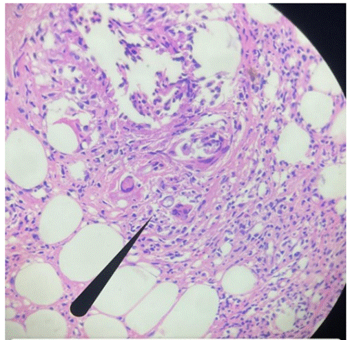
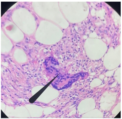
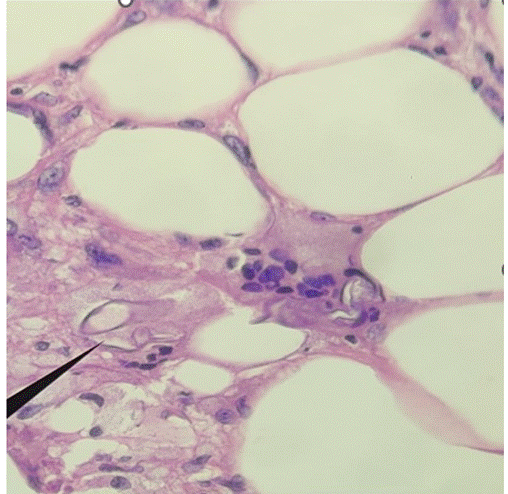
    <heading><span><bold>INTRODUCTION</bold></span></heading><p><span>Granulomatous inflammation is a distinctive form of chronic inflammation produced in response to various infections, autoimmune, toxic, allergic and neoplastic conditions<superscript><superscript>[<xref ref-type="link" rid="#ref-1">1</xref>]</superscript></superscript></span><italic><span>. </span></italic><span>Fungal granulomas are rare, chronic infections with a very low prevalence rate that often resemble tumors or chronic inflammation, complicating diagnosis. While these infections commonly affect immunocompromised individuals, they can also appear in healthy elderly patients, particularly in tropical areas like India. </span></p><p><span>Fungi such as </span><italic><span>Basidiobolus ranarum</span></italic><span>, </span><italic><span>Sporothrix schenckii</span></italic><span>, and </span><italic><span>Madurella mycetomatis</span></italic><span> are frequently seen. A study estimated that over 57 million people suffer from serious fungal diseases<superscript><superscript>[<xref ref-type="link" rid="#ref-2">2</xref>]</superscript></superscript>. Granulomatous invasive fungal sinusitis was seen in nearly 30% of chronic fungal rhinosinusitis cases<superscript><superscript>[<xref ref-type="link" rid="#ref-3">3</xref>]</superscript></superscript>. Globally, more than 6.5 million cases of invasive fungal infections are reported annually, with a significant mortality rate<superscript><superscript>[<xref ref-type="link" rid="#ref-4">4</xref>]</superscript></superscript>. Fungal infections can be difficult to diagnose, although clinicians from various specialties may face this challenge<superscript><superscript>[<xref ref-type="link" rid="#ref-5">5</xref>]</superscript></superscript>.</span></p><p><span>These deep fungal infections can present to the dermatologist with variable presentations, such as nodulo-pustular lesions, cysts, indurated masses with surface changes such as discharging sinuses or verrucosity and ulcer. The lesions generally remain localized and appear as non-tender nodular edema. As a result, they are frequently misidentified as other benign lesions clinically<superscript><superscript>[<xref ref-type="link" rid="#ref-6">6</xref>]</superscript></superscript>. The variable clinical presentation can pose diagnostic challenges<superscript><superscript>[<xref ref-type="link" rid="#ref-7">7</xref>, <xref ref-type="link" rid="#ref-8">8</xref>]</superscript></superscript>. Patients may be treated with topical corticosteroids or calcineurin inhibitors. This may result in tinea incognita, a fungal infection with a different clinical presentation that may complicate the clinician even more<superscript><superscript>[<xref ref-type="link" rid="#ref-9">9</xref>]</superscript></superscript>.</span></p><p><span>Such suspected fungal infections require early evaluation by histopathology, culture aided by imaging studies. Based on which initiation of appropriate therapy to prevent complications.</span></p><heading><span><bold>CASE SUMMARY</bold></span></heading><p><span>This is a clinical case of a 74-year-old female patient came with a complaint of swelling over the right thigh since 3 months. The patient was apparently asymptomatic 3 months back, then developed swelling over the right thigh. The swelling was insidious in onset and gradually progressive, and was associated with pain. The pain was throbbing in nature, increased on walking, and was relieved with medication. No complaints of fever, weight loss, appetite loss, or trauma. She had no known allergies or addictions. There was no history of chronic illness, immunosuppression, or long-term medication use, and has no significant family history. </span></p><p><span>On examination, diffuse swelling is present over the anterior and posterior aspects of the thigh, extending to the distal one-third of the thigh with a size of 10 x 8 x 2cm.  On palpation, tenderness is present over the swelling. No scars, sinus noted over the swelling, no visible pulsations. The provisional diagnosis was soft tissue sarcoma. Clinically Differential Diagnosis considered are Lipoma, Abscess, and Malignancy.</span></p><p><span><bold>Investigations:</bold></span></p><list><list-item><p><span>MRI Pelvis: Hypertrophied, edematous muscles of the right thigh and pelvis.             </span></p></list-item><list-item><p><span>FNAC  of the swelling: Suggestive of a giant cell-containing lesion.</span></p></list-item><list-item><p><span>Hematology: Hb 10.6 g/dL indicates mild anemia, elevated TLC 19,000/mm³, indicating possible infection.</span></p></list-item><list-item><p><span>Histopathology of tissue examined shows dermis and subcutaneous fat, infiltrated with multiple granulomas with necrotic centres. These granulomas are composed of epithelioid cells, foreign-body type giant cells, lymphocytes, and eosinophils <xref ref-type="link" rid="#figure-1">[Fig. 1]</xref>. At places broad, pauci septate, small ribbon-like hyphae are seen in the giant cells and the granulomatous areas <xref ref-type="link" rid="#figure-2">[Fig. 2]</xref> and <xref ref-type="link" rid="#figure-3">[Fig. 3]</xref>. Histopathology diagnosis given as<bold> </bold>Subcutaneous fungal granuloma.</span></p></list-item></list><figure id="figure-1"><graphic src="https://schoproductionportal.s3.ap-south-1.amazonaws.com/data/PIMR/443/1787832857671.png"/></figure><p><span><bold>Fig. 1: Granulomatous lesion containing fungal hyphae (H&amp;E, 10x)</bold></span></p><figure id="figure-2"><graphic src="https://schoproductionportal.s3.ap-south-1.amazonaws.com/data/PIMR/443/1787832857713.png"/></figure><p><span><bold>Fig. 2: Under the light microscopy, fungal hyphae seen in a giant cell (H&amp;E,40x)</bold></span></p><figure id="figure-3"><graphic src="https://schoproductionportal.s3.ap-south-1.amazonaws.com/data/PIMR/443/1787832857652.png"/></figure><p><span><bold>Fig. 3: Fungal hyphae (arrow) (H&amp;E,40x)</bold></span></p><p> </p><p><span><bold>Treatment and follow-up:</bold> </span></p><p><span>Following histopathological diagnosis, the patient was prescribed itraconazole 200 mg orally once daily for 3 days and on follow-up, there was clinical improvement in the swelling and associated pain, and the patient was reported to be clinically well without significant ongoing symptoms. On subsequent follow-up, clinical improvement in the swelling and associated pain was noted, and the patient was clinically well without significant ongoing symptoms.</span></p><heading><span><bold>DISCUSSION</bold></span></heading><p><span>The primary cutaneous fungal pathogens can be classified into two groups:  Superficial Fungal Infections and Deep Fungal Infections. Superficial Fungal Infections are characterized by hyphae or pseudo-hyphae and sometimes yeast cells in the keratin layer of the epidermis and the follicles. Organisms causing superficial cutaneous fungal infections do not affect the dermis, except in cases of hair follicle rupture<superscript><superscript>[<xref ref-type="link" rid="#ref-10">10</xref>]</superscript></superscript>. Deep cutaneous infections affect the dermis and subcutaneous tissue. </span></p><p><span>This case shows fungal hyphae-containing granulomas in deep dermis and subcutaneous fat tissue. The deep fungal infections encountered include chromoblastomycosis, sporotrichosis, rhinosporidiosis, mycetoma, histoplasmosis, subcutaneous pheohyphomycosis, mucor-mycosis, hyalohyphomycosis, entomophthoromycosis, penicilliosis and cryptococcosis<superscript><superscript>[<xref ref-type="link" rid="#ref-11">11</xref>]</superscript></superscript>. This is a case of deep fungal infection involving dermis and subcutaneous fat tissue, characterized by granulomas. The appearance of granulomas in these infections indicates a type IV hypersensitivity response, primarily mediated by CD4+ and CD8+ T-lymphocytes<superscript><superscript>[<xref ref-type="link" rid="#ref-12">12</xref>]</superscript></superscript>.</span></p><p><span>In the differential diagnosis of granulomatous infection of subcutaneous tissue, include cutaneous TB, Crohn’s disease, sarcoidosis, rheumatoid nodules and subcutaneous granuloma annulare. All these are characterized by granulomas, but in our case, along with granulomas, fungal hyphae were also seen, which helped in making the final diagnosis and excluding other granulomatous infections. </span></p><p><span>In the diagnosis of granulomatous inflammation, along with the careful examination of Histopathological sections, use of special stains increases the diagnostic sensitivity. Mycobacterium and fungal infections are the most common cause for granulomatous inflammations, Grocott Methenamine Silver (GMS) stains and Ziehl-Neelsen stain (AFB) are most commonly used special stains for the identification of fungi and acid-fast bacilli. Though culture is the gold standard method for the diagnosis of infectious organism, use molecular diagnostic methods from culture growth or from the fresh or formalin fixed infected tissue improves the specificity and identification of organisms, particularly mycobacteria<superscript><superscript>[<xref ref-type="link" rid="#ref-13">13</xref>, <xref ref-type="link" rid="#ref-14">14</xref>]</superscript></superscript>. <bold> </bold></span></p><heading><span><bold>CONCLUSION</bold></span></heading><p><span>This case highlights the importance of considering subcutaneous fungal infections in the differential diagnosis of chronic soft tissue swelling cases, even in elderly patients without immunosuppression. The histopathological evaluation of granulomatous inflammation is a useful indicator of the diagnosis. A clinical discussion between the clinician and the pathologist enables the appropriate use of necessary laboratory procedures, resulting in accurate and early detection of the disease. Specific therapy helps in the successful care and recovery of the patient without any complications.</span></p>
  </body>
  <back>
    <ref-list>
      <title>References</title>
      
        
          <ref id="ref-1">
            <element-citation publication-type="journal">
              
                <person-group person-group-type="author">
                  <name>
                    Shah KK, Pritt BS, Alexander MP
                  </name>
                </person-group>
              
              
                <article-title>Histopathologic review of granulomatous inflammation</article-title>
              
              
                <source>Journal of Clinical Tuberculosis and Other Mycobacterial Diseases</source>
              
              
                <year>2017</year>
              
              
                <volume>7</volume>


                <uri>https://doi.org/10.1016/j.jctube.2017.02.001</uri>
              
            </element-citation>
          </ref>


          <ref id="ref-2">
            <element-citation publication-type="journal">
              
                <person-group person-group-type="author">
                  <name>
                    Ray A, Aayilliath KA, Banerjee S, Chakrabarti A, Denning DW
                  </name>
                </person-group>
              
              
                <article-title>Burden of Serious Fungal Infections in India</article-title>
              
              
                <source>Open Forum Infectious Diseases</source>
              
              
                <year>2022</year>
              
              
                <volume>9</volume>
              
              
                <issue>12</issue>
              
              
                <uri>https://doi.org/10.1093/ofid/ofac603</uri>
              
            </element-citation>
          </ref>


          <ref id="ref-3">
            <element-citation publication-type="journal">
              
                <person-group person-group-type="author">
                  <name>
                    Sharif MS, Ali S, Nisar H
                  </name>
                </person-group>
              
              
                <article-title>Frequency of Granulomatous Invasive Fungal Sinusitis in Patients with Clinical Suspicion of Chronic Fungal Rhinosinusitis</article-title>
              
              
                <source>Cureus</source>
              
              
                <year>2019</year>
              
              
                <volume>11</volume>
              
              
                <issue>5</issue>
              
              
                <uri>https://doi.org/10.7759/cureus.4757</uri>
              
            </element-citation>
          </ref>


          <ref id="ref-4">
            <element-citation publication-type="journal">
              
                <person-group person-group-type="author">
                  <name>
                    Bongomin F, Gago S, Oladele RO, Denning DW
                  </name>
                </person-group>
              
              
                <article-title>Global and Multi-National Prevalence of Fungal Diseases—Estimate Precision</article-title>
              
              
                <source>Journal of Fungi</source>
              
              
                <year>2017</year>
              
              
                <volume>3</volume>
              
              
                <issue>4</issue>
              
              
                <uri>https://doi.org/10.3390/jof3040057</uri>
              
            </element-citation>
          </ref>


          <ref id="ref-5">
            <element-citation publication-type="journal">
              
                <person-group person-group-type="author">
                  <name>
                    Nowowiejska J, Baran A, Flisiak I
                  </name>
                </person-group>
              
              
                <article-title>Tinea Incognito—A Great Physician Pitfall</article-title>
              
              
                <source>Journal of Fungi</source>
              
              
                <year>2022</year>
              
              
                <volume>8</volume>
              
              
                <issue>3</issue>
              
              
                <uri>https://doi.org/10.3390/jof8030312</uri>
              
            </element-citation>
          </ref>


          <ref id="ref-6">
            <element-citation publication-type="journal">
              
                <person-group person-group-type="author">
                  <name>
                    Priyadharshini G, Varghese RG, Phansalkar M, Ramdas A
                  </name>
                </person-group>
              
              
                <article-title>Subcutaneous Fungal Cyst Masquerading as Benign Lesions – A Series of Eight Cases</article-title>
              
              
                <source>Journal of Clinical and Diagnostic Research</source>
              
              
                <year>2015</year>
              
              
                <volume>9</volume>
              
              
                <issue>10</issue>
              
              
                <uri>https://doi.org/10.7860/jcdr/2015/14157.6637</uri>
              
            </element-citation>
          </ref>


          <ref id="ref-7">
            <element-citation publication-type="journal">
              
                <person-group person-group-type="author">
                  <name>
                    Verma S, Thakur BK, Raphael V, Thappa DM
                  </name>
                </person-group>
              
              
                <article-title>Epidemiology of subcutaneous mycoses in Northeast India: a retrospective study</article-title>
              
              
                <source>Indian Journal of Dermatology</source>
              
              
                <year>2018</year>
              
              
                <volume>63</volume>
              
              
                <issue>6</issue>
              
              
                <uri>https://doi.org/10.4103/ijd.IJD_16_18</uri>
              
            </element-citation>
          </ref>


          <ref id="ref-8">
            <element-citation publication-type="journal">
              
                <person-group person-group-type="author">
                  <name>
                    Samaila MO, Abdullahi K
                  </name>
                </person-group>
              
              
                <article-title>Cutaneous manifestations of deep mycosis: an experience in a tropical pathology laboratory</article-title>
              
              
                <source>Indian Journal of Dermatology</source>
              
              
                <year>2011</year>
              
              
                <volume>56</volume>
              
              
                <issue>3</issue>
              
              
                <uri>https://doi.org/10.4103/0019-5154.82481</uri>
              
            </element-citation>
          </ref>


          <ref id="ref-9">
            <element-citation publication-type="journal">
              
                <person-group person-group-type="author">
                  <name>
                    Drivenes JL, Ramsing M, Bygum A
                  </name>
                </person-group>
              
              
                <article-title>Majocchi’s Granuloma – The Great Mimicker: A Case Report</article-title>
              
              
                <source>Case Reports in Dermatology</source>
              
              
                <year>2023</year>
              
              
                <volume>15</volume>
              
              
                <issue>1</issue>
              
              
                <uri>https://doi.org/10.1159/000533475</uri>
              
            </element-citation>
          </ref>


          <ref id="ref-10">
            <element-citation publication-type="journal">
              
                <person-group person-group-type="author">
                  <name>
                    Hinshaw M, Longley BJ
                  </name>
                </person-group>
              
              
                <article-title>Fungal Diseases. In: Elder DE, Elenitsas R, Murphy GF, Johnson Jr BL, Xu X</article-title>
              
              
                <source>&lt;I&gt;Lever’s Histopathology of skin&lt;/I&gt;</source>
              
              
                <year>2011</year>
              
              
                <volume>10th ed</volume>


                <uri>https://books.google.co.in/books/about/Lever_s_Histopathology_of_the_Skin.html?id=R3pMLydb1NMC&amp;redir_esc=y</uri>
              
            </element-citation>
          </ref>


          <ref id="ref-11">
            <element-citation publication-type="journal">
              
                <person-group person-group-type="author">
                  <name>
                    Dhar S, Pradhan S, Saha A, Mazumder G, Ghosh SK, Biswas M, &lt;I&gt;et al&lt;/I&gt;
                  </name>
                </person-group>
              
              
                <article-title>Deep Fungal Infections of Skin and Role of Histopathology in Diagnosis</article-title>
              
              
                <source>Indian Journal of Dermatology</source>
              
              
                <year>2024</year>
              
              
                <volume>69</volume>
              
              
                <issue>6</issue>
              
              
                <uri>https://doi.org/10.4103/ijd.ijd_419_23</uri>
              
            </element-citation>
          </ref>


          <ref id="ref-12">
            <element-citation publication-type="journal">
              
                <person-group person-group-type="author">
                  <name>
                    Badea MA, Buicu CF, Sin AI, Cotoi OS, Badea IM, Chiotoroiu AL, &lt;I&gt;et al&lt;/I&gt;
                  </name>
                </person-group>
              
              
                <article-title>The value of histopathological diagnosis in the elderly patients with granulomatous dermatoses. Case series</article-title>
              
              
                <source>Romanian Journal of Morphology and Embryology</source>
              
              
                <year>2016</year>
              
              
                <volume>57</volume>
              
              
                <issue>2</issue>
              
              
                <uri>https://pubmed.ncbi.nlm.nih.gov/27516029/</uri>
              
            </element-citation>
          </ref>


          <ref id="ref-13">
            <element-citation publication-type="journal">
              
                <person-group person-group-type="author">
                  <name>
                    Kadhirvelu HK, Murugan S, Ramakrishnan R, Premamalini T
                  </name>
                </person-group>
              
              
                <article-title>Subcutaneous zygomycosis in an immunocompetent patient caused by &lt;i&gt;Basidiobolus ranarum&lt;/i&gt;</article-title>
              
              
                <source>BMJ Case Reports</source>
              
              
                <year>2023</year>
              
              
                <volume>16</volume>
              
              
                <issue>1</issue>
              
              
                <uri>https://doi.org/10.1136/bcr-2022-253515</uri>
              
            </element-citation>
          </ref>


          <ref id="ref-14">
            <element-citation publication-type="journal">
              
                <person-group person-group-type="author">
                  <name>
                    Caulfield AJ, Wengenack NL
                  </name>
                </person-group>
              
              
                <article-title>Diagnosis of active tuberculosis disease: From microscopy to molecular techniques</article-title>
              
              
                <source>Journal of Clinical Tuberculosis and Other Mycobacterial Diseases</source>
              
              
                <year>2016</year>
              
              
                <volume>4</volume>


                <uri>https://doi.org/10.1016/j.jctube.2016.05.005</uri>
              
            </element-citation>
          </ref>
        
      
    </ref-list>
  </back>
</article>