Volume: 14 Issue: 1
Effect of Guided Imagery on Occupational Therapy Functional Outcomes in Acute Burns Care.
Year: 2026, Page: 46-51, Doi: https://doi.org/10.47799/pimr.1401.26.40
Received: March 30, 2026 Accepted: April 16, 2026 Published: May 2, 2026
Abstract
Burn injuries constitute a major global health problem and frequently lead to severe pain, anxiety, functional limitations, and long-term physical and psychological complications. Inadequate pain control and anticipatory anxiety during repeated procedures often hinder rehabilitation participation and delay functional recovery. Guided imagery, a mind–body intervention, has demonstrated potential in reducing pain perception and promoting relaxation by influencing autonomic and neuroendocrine responses. This study examined the efficacy of guided imagery as an adjunct to occupational therapy (OT) in acute burns care. A quasi-experimental study was conducted with 40 patients with acute burns recruited through convenience sampling and randomly allocated into two groups (n = 20 each). The control group received routine OT exercises, while the experimental group received guided imagery in addition to routine OT for 45 minutes, twice weekly, over four weeks. Pain was assessed using the Visual Analogue Scale (VAS), anxiety using the Hamilton Anxiety Rating Scale (HAM-A), and functional limitation using the Patient-Specific Functional Scale (PSFS). Pulse rate and respiratory rate were measured before and after intervention. Both groups demonstrated significant improvement in pain (VAS, p < 0.01) and vital signs (pulse and respiratory rate, p < 0.01). However, no significant changes were observed in anxiety (HAM-A) or functional performance (PSFS). The findings suggest that guided imagery serves as a beneficial adjunct to occupational therapy in reducing pain and stabilizing physiological responses in patients with acute burns.
Keywords: Acute burns, Anxiety, Guided imagery, Occupational therapy, Pain
INTRODUCTION
Burn injuries continue to be a significant health problem worldwide and are associated with considerable morbidity[1]. Patients with burns often experience severe pain, especially during procedures such as dressing changes and physical therapy[1, 2]. This pain is not only physical but also influenced by anxiety and fear related to repeated interventions[3]. In the acute phase, inflammatory pain is more prominent, and later it may get complicated by neuropathic components[1, 4]. Poorly controlled pain can delay recovery and reduce participation in rehabilitation[2, 5].
Apart from pain, patients with burns commonly develop psychological issues such as anxiety, distress, and fear of movement[3]. These factors can interfere with engagement in therapy and may affect long-term functional outcomes[3, 5]. Even though pharmacological management, including opioids and sedatives, is routinely used, complete relief is often not achieved and side effects remain a concern[2]. Because of this, there is increasing interest in non-pharmacological methods which can be used along with routine care[6].
Various approaches such as virtual reality, relaxation techniques, and guided imagery have been studied for pain management in burn patients[6, 7]. Guided imagery is a simple technique in which the patient is encouraged to form mental images that help in relaxation and distraction from pain[7]. It is believed to act through cognitive and emotional pathways and may influence the perception of pain[6, 8]. Some studies have shown that guided imagery can reduce pain and anxiety in different clinical settings, including surgical procedures and chronic conditions[8-10].
In burn care, occupational therapy plays an important role in improving functional ability and preventing long-term disability. Early mobilisation, splinting, and functional training are essential components of rehabilitation[5, 11]. However, pain and anxiety during therapy sessions can reduce patient cooperation and limit the effectiveness of these interventions[2, 3]. Techniques that can improve patient comfort may help in better participation and outcomes[5].
Few studies have examined the use of guided imagery specifically in burn patients during rehabilitation. Aghakhani et al. reported reduction in pain and anxiety during dressing procedures with guided imagery[12]. Similar findings have been reported with relaxation and imagery-based techniques in burn and non-burn populations[13-15]. However, there is limited evidence regarding its role when combined with occupational therapy in the acute phase of burn care[5, 12].
Considering this gap, the present study was undertaken to evaluate the effect of guided imagery as an adjunct to occupational therapy on pain, anxiety, and functional outcomes in patients with acute burns.
MATERIALS AND METHODS
Study Design and Setting: This was a prospective, interventional, assessor-blinded, randomised controlled study conducted in the acute burns unit and occupational therapy department of a tertiary care hospital in India. The study protocol was approved by the institutional ethics committee (ECARP/2021/104) and written informed consent was obtained from all participants prior to enrolment.
Sample Size: A convenience sample of 40 participants was recruited. Sample size estimation was based on a prior study by Aghakhani et al., which reported a mean difference in pain scores of 2.1 (SD ± 1.8) between guided imagery and control groups in burn patients[12]. Using a two-tailed independent t-test with α = 0.05 and power (1 − β) = 0.80, a minimum of 18 participants per group was required. Accounting for a 10% dropout, 20 participants per group were enrolled.
Participants: Patients aged 18 years and above with acute, non-inhalational, non-intentional first or second degree burn injuries who were haemodynamically stable and had a pain score of ≥ 5 on the Visual Analogue Scale were eligible for inclusion. Patients admitted to the intensive care unit or those with a pre-existing diagnosed psychiatric illness were excluded from the study.
Recruitment and Allocation: Eligible participants were recruited by convenience sampling from patients referred to the occupational therapy department from the acute burns unit. Following enrolment, allocation to groups was performed using a simple lottery method, in which sealed cards marked 'A' (Control) and 'B' (Experimental) were prepared in equal numbers (n = 20 each) and drawn by each participant. Due to the nature of the guided imagery intervention, blinding of participants was not feasible; however, all primary and secondary outcome assessments were performed by an independent assessor who was not involved in intervention delivery, thereby ensuring assessor blinding.
All participants continued to receive standard pharmacological management for burn pain as per institutional protocol under the supervision of the treating surgical team. The occupational therapy team did not modify analgesic prescriptions during the study period, and no additional pharmacological intervention specific to either group was introduced.
Intervention: Participants in the control group (Group A) received routine occupational therapy, which comprised range-of-motion exercises, functional task training, use of adaptive devices, splinting where clinically indicated, and a graduated programme of dexterity activities. Pressure garments were prescribed once adequate tissue healing had occurred, and pre-discharge activities targeting fine motor function and flexibility were incorporated as appropriate. Each session was supervised by a trained occupational therapist.
Participants in the experimental group (Group B) received the same routine occupational therapy programme in addition to a structured guided imagery intervention. Sessions were conducted for 45 minutes each, twice weekly, over a period of four weeks (total: eight sessions). The guided imagery protocol consisted of two sequential phases. In the first phase, a relaxation response was induced through controlled breathing exercises followed by progressive muscle relaxation, proceeding systematically from distal to proximal muscle groups. In the second phase, participants were guided to form a mental image of their pain and, with facilitator guidance, to transform that image into a less distressing or neutral representation ([Fig. 1] & [Fig. 2]). The procedure was explained to each participant prior to commencement, and verbal confirmation of understanding was obtained.
Outcome Measures: All outcome measures were assessed at baseline (pre-intervention) and at the end of four weeks (post-intervention) by the independent assessor.
Pain intensity was assessed using the Visual Analogue Scale (VAS), a 100 mm horizontal line on which 0 indicates no pain and 10 indicates the worst imaginable pain[26]. Anxiety was assessed using the Hamilton Anxiety Rating Scale (HAM-A), a clinician-administered scale comprising 14 items each rated 0–4, with higher total scores indicating greater anxiety severity[17]. Functional status was assessed using the Patient-Specific Functional Scale (PSFS), on which participants rate their ability to perform up to five self-selected functional activities on an 11-point scale from 0 (unable to perform) to 10 (able to perform at prior level); higher scores indicate better functional ability[16, 18].
Pulse rate (PR) and respiratory rate (RR) were recorded as secondary physiological outcome measures. These were measured immediately before and after each intervention session throughout the four-week period, and the pre- and post-intervention values recorded at weeks 0 and 4 were used for statistical analysis.
Statistical Analysis: Data were analysed using IBM SPSS Statistics version 21.0 (IBM Corp., Armonk, NY, USA). Continuous variables are presented as mean ± standard deviation (SD), and categorical variables as frequency and percentage. Normality of continuous data was assessed using the Shapiro–Wilk test. Appropriate non-parametric tests were used for all comparisons (Intra- and inter-groups) of data not normally distributed. A p value of less than 0.05 was considered statistically significant.
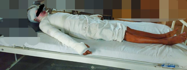
Fig. 1: Guided imagery intervention
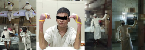
Fig. 2: Activities for hand dexterity with complex motion by using pegs of different shape and sizes, adaptive device and functional activities
RESULTS
Baseline characteristics: A total of 40 participants were included, with 20 patients in each group. The groups were comparable with respect to age, gender distribution, type of burn, total body surface area [Table. 1]. Concomitant treatment variables, including pharmacological analgesic use, were similar across both groups.
| Variables | Category | Experimental (n=20) | Control (n=20) | p value |
|---|---|---|---|---|
| Age (years) | Mean ± SD | 37.35 ± 14.80 | 35.70 ± 13.70 | t = 0.366, p=0.717 |
| Gender | Male | 10 | 13 | χ² = 0.921, p=0.337 |
| Female | 10 | 7 | ||
| Type of burn | Flame | 13 | 10 | χ² = 3.12, p=0.537 |
| Electrical | 2 | 5 | ||
| Scald | 2 | 3 | ||
| Chemical | 2 | 2 | ||
| Acid | 1 | 0 | ||
| TBSA (%) | Mean ± SD | 30.6 ± 8.39 | 29.9 ± 9.65 | U = 186.5, p=0.713 |
|
PSFS (baseline) |
Mean ± SD | 4.00 ± 1.26 | 3.15 ± 1.27 | U = 143.0, p=0.083 |
Table 1: Baseline Characteristics of Participants in Experimental and Control Groups
TBSA = Total Body Surface Area, PSFS = Patient Specific Functional Scale.
Intra-group Analysis: Pre- to Post-Intervention Changes
Both groups showed significant improvement in all outcome measures following four weeks of intervention [Table. 2]. Pain intensity as measured by the VAS, and Anxiety scores on the HAM-A decreased significantly in both the experimental and control groups. Functional status improved significantly, as reflected by increased Patient Specific Functional Scale scores. Pulse rate and respiratory rate demonstrated significant reduction in both groups over the study period.
| Outcome | Group | Pre-Intervention (Mean ± SD) |
Post-Intervention (Mean ± SD) |
Z value | p value |
|---|---|---|---|---|---|
| VAS | Experimental | 8.1 ± 0.85 | 0.7 ± 0.86 | -4.018 | <0.001 |
| Control | 7.9 ± 0.85 | 1.6 ± 0.94 | -4.099 | <0.001 | |
| HAM-A | Experimental | 31.1 ± 11.48 | 2.05 ± 2.16 | -3.928 | <0.001 |
| Control | 29.4 ± 10.23 | 3.00 ± 1.75 | -3.924 | <0.001 | |
| PSFS | Experimental | 4.00 ± 1.26 | 9.45 ± 0.61 | -3.970 | <0.001 |
| Control | 3.15 ± 1.27 | 8.25 ± 1.37 | -3.945 | <0.001 | |
| Pulse rate | Experimental | 84.7 ± 7.09 | 76.5 ± 4.98 | -3.980 | <0.001 |
| Control | 87.7 ± 5.99 | 82.7 ± 6.72 | -3.955 | <0.001 | |
| Respiratory rate | Experimental | 28.1 ± 1.92 | 17.8 ± 1.58 | -3.946 | <0.001 |
| Control | 26.65 ± 3.59 | 19.2 ± 2.09 | -3.943 | <0.001 |
Table 2: Intra-group comparison of outcome measures at baseline and after four weeks of intervention
VAS = Visual Analogue Scale; HAM-A = Hamilton Anxiety Rating Scale; PSFS = Patient-Specific Functional Scale. *p < 0.001, statistically significant by Wilcoxon signed-rank test. Values are mean ± SD.
Inter-group Analysis: Between-Group Comparison of Change Scores
Between-group comparisons of change scores (post minus pre) using the Mann–Whitney U test are summarised in [Table. 3]. The reduction in pain (VAS change score) was significantly greater in the experimental group (7.4 ± 0.68) compared with the control group (6.3 ± 0.47), p<0.001 with a large effect size. Reduction in pulse rate and respiratory rate was also significantly greater in the experimental group.
There was no statistically significant difference between groups for anxiety (HAM-A, p = 0.355; r = 0.15) or functional status (PSFS, p = 0.552; r = 0.09), indicating comparable improvement in these outcomes.
| Outcome (Change Score) | Experimental Mean ± SD |
Control Mean ± SD |
U value | p value | Effect Size (r) |
|---|---|---|---|---|---|
| VAS (pain) | 7.4 ± 0.68 | 6.3 ± 0.47 | 46 | < 0.001* | 0.71 |
| HAM-A (anxiety) | 29.05 ± 9.51 | 26.4 ± 8.97 | 166 | 0.355 | 0.15 |
| PSFS (function) | 5.35 ± 1.46 | 5.1 ± 1.68 | 179 | 0.552 | 0.09 |
| Pulse rate (bpm) | 8.2 ± 2.89 | 5.0 ± 2.2 | 71.5 | < 0.001* | 0.57 |
| Respiratory rate (breaths/min) | 10.3 ± 2.05 | 7.45 ± 2.46 | 78.5 | 0.001* | 0.53 |
Table 3: Inter-group comparison of change scores between experimental and control groups
VAS = Visual Analogue Scale; HAM-A = Hamilton Anxiety Rating Scale; PSFS = Patient-Specific Functional Scale; bpm = beats per minute. *p < 0.05, statistically significant. Effect size r derived from rank-biserial correlation: 0.1 = small, 0.3 = medium, 0.5 = large. Values are mean ± SD.
DISCUSSION
The present study evaluated the effect of guided imagery as an adjunct to occupational therapy in patients with acute burns. Both groups showed significant improvement in pain, anxiety, functional status, and physiological parameters over the study period. This indicates that routine occupational therapy along with standard medical management contributes substantially to recovery in burn patients, as reported in earlier rehabilitation studies[19-22].
A greater reduction in pain was observed in the experimental group as compared to the control group. This is consistent with previous studies in burn care. Aghakhani et al. reported reduction in pain during dressing procedures with guided imagery[12]. Similar findings have been reported with guided imagery and relaxation-based techniques in other clinical settings[10, 23]. These interventions are thought to reduce pain perception through cognitive and emotional mechanisms. Other non-pharmacological approaches such as relaxation, distraction, and virtual reality have also shown benefit in reducing pain[6, 7, 13]. These methods are particularly useful when pharmacological measures alone are not sufficient[2].
In the present study, anxiety improved in both groups, but no additional benefit of guided imagery was observed. This finding is in agreement with some previous studies where no significant difference was observed when guided imagery was added to routine care[14]. It is possible that factors such as repeated exposure to therapy, interaction with healthcare staff, and structured rehabilitation environment contribute to reduction in anxiety. Basic relaxation responses may also play a role in this improvement[24].
Functional outcomes improved significantly in both groups, but there was no difference between groups. This suggests that functional recovery is mainly driven by occupational therapy interventions such as mobilization, splinting, and task-oriented training[5, 11]. Similar observations have been reported in burn rehabilitation studies where early and consistent therapy was the key determinant of functional improvement[19-22]. Guided imagery may help in symptom relief but does not appear to influence functional outcomes over a short duration.
The reduction in pulse rate and respiratory rate was greater in the experimental group. This supports the physiological effect of guided imagery in reducing autonomic arousal. Relaxation-based interventions are known to reduce sympathetic activity and promote parasympathetic response, which may explain these findings[15, 24, 25].
The findings of this study should be interpreted with certain limitations. The sample size was small, and participants were selected by convenience sampling, which may limit generalisability. The study was conducted in a single centre with short follow up. Although pharmacological management was similar across groups, detailed quantification of analgesic use was not performed. Further studies with larger sample size and longer follow up are required.
CONCLUSION
The findings of the present study indicate that guided imagery, when incorporated as an adjunct to routine occupational therapy in patients with acute burns, results in a significantly greater reduction in pain and improvement in physiological parameters such as pulse and respiratory rate. However, no additional advantage was observed with respect to anxiety reduction or functional recovery when compared to routine therapy alone. These observations suggest that while guided imagery may have a role in enhancing pain control during the acute rehabilitation phase, its influence on psychological and functional outcomes appears limited over the short term. In contrast, functional improvement continues to be largely determined by structured occupational therapy interventions. Given the methodological limitations and short duration of follow-up, further well-designed studies with larger sample sizes are required to establish the clinical utility and long-term impact of guided imagery in burn rehabilitation.
DISCLOSURE
Funding: Nil.
Conflict of Interest: None.
References
1. Summer GJ, Puntillo KA, Miaskowski C, Green PG, Levine JD. Burn Injury Pain: The Continuing Challenge. The Journal of Pain. 2007; 8 (7). Available from: https://doi.org/10.1016/j.jpain.2007.02.426
2. Griggs C, Goverman J, Bittner EA, Levi B. Sedation and Pain Management in Burn Patients. Clinics in Plastic Surgery. 2017; 44 (3). Available from: https://doi.org/10.1016/j.cps.2017.02.026
3. Van Loey NE, Van Son MJ. Psychopathology and Psychological Problems in Patients with Burn Scars. American Journal of Clinical Dermatology. 2003; 4 (4). Available from: https://doi.org/10.2165/00128071-200304040-00004
4. Morad A, Farrokh S. Pain management. Essentials of Anesthesia for Neurotrauma. 2018; Available from: https://doi.org/10.1201/9781315166742-32
5. Aghajanzade M, Momeni M, Niazi M, Ghorbani H, Saberi M, Kheirkhah R, et al. Effectiveness of incorporating occupational therapy in rehabilitation of hand burn patients. Annals of Burns and Fire Disasters. 2019;32(2):147–152.
6. Sharar SR, Miller W, Teeley A, Soltani M, Hoffman HG, Jensen MP, et al. Applications of virtual reality for pain management in burn-injured patients. Expert Review of Neurotherapeutics. 2008; 8 (11). Available from: https://doi.org/10.1586/14737175.8.11.1667
7. Bonadies V. Guided imagery as a therapeutic recreation modality to reduce pain and anxiety. Ther Recreat J. 2009;43(2):43–52.
8. Forward JB, Greuter NE, Crisall SJ, Lester HF. Effect of Structured Touch and Guided Imagery for Pain and Anxiety in Elective Joint Replacement Patients—A Randomized Controlled Trial: M-TIJRP. The Permanente Journal. 2015; 19 (4). Available from: https://doi.org/10.7812/tpp/14-236
9. Gonzales EA, Ledesma RJ, McAllister DJ, Perry SM, Dyer CA, Maye JP. Effects of guided imagery on postoperative outcomes in patients undergoing same-day surgical procedures: a randomized single-blind study. AANA Journal. 2010 Jun;78(3):181-8. PMID: 20572403.
10. Baird CL, Sands L. A pilot study of the effectiveness of guided imagery with progressive muscle relaxation to reduce chronic pain and mobility difficulties of osteoarthritis. Pain Management Nursing. 2004; 5 (3). Available from: https://doi.org/10.1016/j.pmn.2004.01.003
11. Pedretti LW, Early MB, editors. Occupational therapy: practice skills for physical dysfunction. 5th ed. St Louis: Mosby; 2001.
12. Aghakhani N, Faraji N, Alinejad V, Goli R, Kazemzadeh J. The effect of guided imagery on the quality and severity of pain and pain-related anxiety associated with dressing changes in burn patients: A randomized controlled trial. Burns. 2022; 48 (6). Available from: https://doi.org/10.1016/j.burns.2021.11.020
13. Delfani F, Zakeri-Moghadam M, Aliha JM. Effect of muscle relaxation and mental imagery techniques on pain in patients with burns. Iranian Journal of Nursing and Midwifery Research. 2016;21(3):256–261.
14. Charalambous A, Giannakopoulou M, Bozas E, Paikousis L. A Randomized Controlled Trial for the Effectiveness of Progressive Muscle Relaxation and Guided Imagery as Anxiety Reducing Interventions in Breast and Prostate Cancer Patients Undergoing Chemotherapy. Evidence-Based Complementary and Alternative Medicine. 2015; 2015 Available from: https://doi.org/10.1155/2015/270876
15. Beizaee Y, Rejeh N, Heravi-Karimooi M, Tadrisi SD, Griffiths P, Vaismoradi M. The effect of guided imagery on anxiety, depression and vital signs in patients on hemodialysis. Complementary Therapies in Clinical Practice. 2018; 33 Available from: https://doi.org/10.1016/j.ctcp.2018.10.008
16. Westaway MD, Stratford PW, Binkley JM. The Patient-Specific Functional Scale: Validation of Its Use in Persons With Neck Dysfunction. Journal of Orthopaedic & Sports Physical Therapy. 1998; 27 (5). Available from: https://doi.org/10.2519/jospt.1998.27.5.331
17. Thompson E. Hamilton Rating Scale for Anxiety (HAM-A). Occupational Medicine. 2015; 65 (7). Available from: https://doi.org/10.1093/occmed/kqv054
18. Moore HE, Corning WL, van der Esch M, Roorda LD, Dekker J, Groot J, et al. Evaluation of treatment outcome using the Patient Specific Functional Scale in knee osteoarthritis patients undergoing multidisciplinary rehabilitation. Osteoarthritis and Cartilage Open. 2020; 2 (4). Available from: https://doi.org/10.1016/j.ocarto.2020.100098
19. Elsherbiny OE, El Fahar MH, Weheida SM, Shebl AM, Shrief WI. Effect of burn rehabilitation program on improving quality of life (QoL) for hand burns patients: a randomized controlled study. European Journal of Plastic Surgery. 2018; 41 (4). Available from: https://doi.org/10.1007/s00238-017-1379-7
20. Spires MC, Bowden ML, Ahrns KS, Wahl WL. Impact of an Inpatient Rehabilitation Facility on Functional Outcome and Length of Stay of Burn Survivors. Journal of Burn Care & Rehabilitation. 2005; 26 (6). Available from: https://doi.org/10.1097/01.bcr.0000185397.39029.0a
21. Tang D, Li-Tsang CW, Au RK, Li KC, Yi XF, Liao LR, et al. Functional Outcomes of Burn Patients with or without Rehabilitation in Mainland China. Hong Kong Journal of Occupational Therapy. 2015; 26 (1). Available from: https://doi.org/10.1016/j.hkjot.2015.08.003
22. DeSanti L, Lincoln L, Egan F, Demling R. Development of a Burn Rehabilitation Unit: Impact on Burn Center Length of Stay and Functional Outcome. Journal of Burn Care & Rehabilitation. 1998; 19 (5). Available from: https://doi.org/10.1097/00004630-199809000-00011
23. Billquist EJ, Michelfelder A, Brincat C, Brubaker L, Fitzgerald CM, Mueller ER. Pre-operative guided imagery in female pelvic medicine and reconstructive surgery: a randomized trial. International Urogynecology Journal. 2018; 29 (8). Available from: https://doi.org/10.1007/s00192-017-3443-z
24. Dusek JA, Benson H. Mind-body medicine: a model of the comparative clinical impact of the acute stress and relaxation responses. Minnesota medicine. 2009 May;92(5):47.
25. Varvani FP, Hekmatpou D, Shamsi KS. Effect of muscle relaxation on burn patients. Iranian Journal of Critical Care Nursing. 2013;6(2):87–94.
26. Wewers ME, Lowe NK. A critical review of visual analogue scales in the measurement of clinical phenomena. Research in Nursing & Health. 1990; 13 (4). Available from: https://doi.org/10.1002/nur.4770130405
Copyright
©2026 (Sawant & Koutarapu). This is an open-access journal, and articles are distributed under the terms of the Creative Commons Attribution License CC-BY 4.0. (https://creativecommons.org/licenses/by/4.0/) which permits unrestricted use, distribution, and reproduction in any medium, provided the original authors and source are credited.
Cite this article
Sawant A, Koutarapu S. Effect of Guided Imagery on Occupational Therapy Functional Outcomes in Acute Burns Care. Perspectives in Medical Research 2026; 14(1):46-51 DOI: 10.47799/pimr.1401.26.40