Volume: 14 Issue: 2
Mixed Epithelial and Stromal Tumor of the Kidney in a Young Male: Report of a Rare Case and Diagnostic Considerations
Year: 2026, Page: 96-99, Doi: https://doi.org/10.47799/pimr.1402.26.62
Received: May 15, 2026 Accepted: June 15, 2026 Published: Aug. 31, 2026
Abstract
Mixed epithelial and stromal tumor (MEST) of the kidney is a rare, predominantly benign biphasic neoplasm, classically seen in perimenopausal women and often associated with estrogen exposure. Occurrence in young males without hormonal therapy is exceptionally uncommon and poses significant diagnostic challenges. We report a case of a 34-year-old male who presented with abdominal discomfort and was found on ultrasonography to have a large retroperitoneal cystic mass, initially misinterpreted as a pancreatic pseudocyst. Subsequent contrast-enhanced CT revealed a mixed solid–cystic lesion arising from the left kidney, radiologically suspicious for malignancy, leading to radical nephrectomy. Histopathological examination demonstrated a characteristic biphasic tumor composed of benign epithelial elements and ovarian-type stromal proliferation. Immunohistochemistry confirmed renal epithelial differentiation and stromal lineage, establishing the diagnosis of renal MEST. The patient remains asymptomatic with no recurrence on follow-up. This case highlights the radiologic pitfalls associated with cystic renal masses, particularly in atypical clinical settings, and emphasizes the pivotal role of histopathology and immunohistochemistry in achieving accurate diagnosis and avoiding potential overtreatment.
Keywords: Mixed epithelial stromal tumor, Kidney, Cystic renal mass, Radiologic–pathologic correlation, Young male
INTRODUCTION
Mixed epithelial and stromal tumor (MEST) is a rare, predominantly benign renal neoplasm, accounting for approximately 0.2% of all renal tumors. In the 2022 World Health Organization (WHO) classification, MEST is classified within the mixed epithelial and stromal tumor family of the kidney, representing a morphologic continuum ranging from predominantly cystic adult cystic nephroma to variably solid and cystic MEST[1]. Adult cystic nephroma, previously regarded as a distinct entity, is now unified with MEST based on overlapping morphologic, immunohistochemical, and molecular features[2]. Histologically, MEST is a biphasic tumor composed of epithelial elements forming glands and cysts embedded within a characteristic ovarian-type stromal background[3].
MEST predominantly affects perimenopausal women and has been associated with estrogenic stimulation, including long-term hormone therapy. Most cases are detected incidentally, although some patients present with nonspecific symptoms such as flank pain, abdominal mass, or hematuria. Occurrence in males is exceedingly rare and has most often been reported in association with prior estrogen exposure[4].
Radiologic diagnosis of MEST remains challenging due to its heterogeneous cystic and solid components and the absence of pathognomonic imaging features. Depending on size, architecture, and growth pattern, MEST may mimic cystic renal cell carcinoma, multilocular cystic renal neoplasm, adult cystic nephroma, or even extrarenal retroperitoneal lesions, frequently leading to radiologic misclassification and potential overtreatment[5].
We report an exceptionally rare case of renal MEST in a young male without any history of hormonal therapy, presenting as a large retroperitoneal cystic mass and posing a significant radiologic diagnostic dilemma. Sequential imaging interpretations included pancreatic pseudocyst and malignant renal neoplasm. This case highlights the critical role of histopathology and immunohistochemistry in establishing the correct diagnosis and underscores the limitations of radiologic evaluation in atypical clinical settings.
CASE REPORT
A 34-year-old male presented with abdominal pain, postprandial fullness, and bloating of 3–4 months’ duration. There was no significant past medical or family history, and no history of hormonal therapy. Physical examination was unremarkable.
Ultrasonography of the abdomen revealed a large retroperitoneal cystic lesion measuring 181.4 × 129.3 × 128.6 mm (volume: 1583.7 cc), containing internal echoes, thick septations, and eccentrically placed solid components. The lesion abutted the body and tail of the pancreas, leading to an initial radiologic impression of pancreatic pseudocyst.
Based on this interpretation, the patient underwent surgical exploration without prior cross-sectional imaging. Complete excision was not feasible intraoperatively, and tissue labelled as “retroperitoneal cyst wall” was submitted for histopathologic examination. Owing to inconclusive intraoperative findings, further radiologic evaluation was performed. Contrast-enhanced computed tomography demonstrated a heterogeneous, moderately enhancing mixed solid–cystic mass arising from the lower pole of the left kidney, radiologically suspicious for a malignant renal neoplasm, particularly renal cell carcinoma [Fig. 1].
During this interval, the patient developed acute clinical deterioration with a sudden decline in haemoglobin levels, likely due to internal haemorrhage from the residual mass.
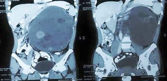
Fig. 1: Contrast-enhanced computed tomography demonstrated a heterogeneous, moderately enhancing mixed solid–cystic mass arising from the lower pole of the left kidney
Following hemodynamic stabilization with blood transfusion, emergency surgery was performed, and a left radical nephrectomy was undertaken in view of strong radiologic suspicion of malignancy.
Histopathologic examination of the previously submitted retroperitoneal cyst wall revealed a biphasic neoplasm composed of epithelial and stromal components. The epithelial component consisted of glands lined by low cuboidal to columnar cells with eosinophilic cytoplasm. The stromal component was cellular, displaying a wavy and whorled architecture reminiscent of ovarian-type stroma, with septal thickness exceeding 5 mm. No cytologic atypia, mitotic activity, or necrosis was identified [Fig. 2].
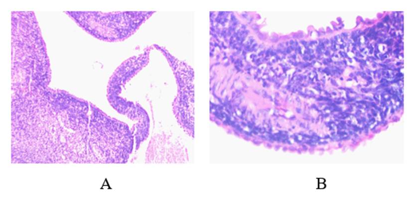
Fig. 2. A) Histopathological examination (H and E stain) showed cyst wall having both epithelial and stromal components (100x). B) Epithelial component showing hobnailing with underlying dense ovarian type stroma (200X)
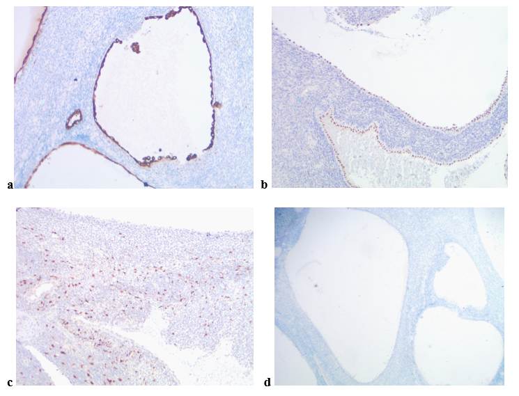
Fig. 3: Immunohistochemical expression of MEST. a and b showing positivity of Pan CK and Pax 8 respectively in epithelial component. c) Focal expression of Calretinin in stroma. d) ER negativity in stroma
Immunohistochemical studies showed positivity for PAX8 and pan-cytokeratin in epithelial elements, with focal CD10 expression [Fig. 3]. Stromal cells demonstrated focal positivity for desmin and calretinin. SMA, HMB45, ER, PR, inhibin, and WT1 were negative in both components. These findings supported a diagnosis of renal mixed epithelial and stromal tumor.
Gross examination of the nephrectomy specimen revealed a cystic lesion arising from the mid-pole of the kidney, with unremarkable upper and lower poles. Histologic features were identical to those seen in the earlier specimen, confirming the diagnosis. Adjacent renal parenchyma was largely unremarkable, apart from mild interstitial lymphoplasmacytic infiltration.
The patient remains asymptomatic with no evidence of recurrence or metastasis on follow-up.
DISCUSSION
MEST is a rare biphasic renal neoplasm classified within the mixed epithelial and stromal tumor family in the WHO 2022 classification, representing a morphologic spectrum that includes adult cystic nephroma[1]. Molecular and clinicopathologic studies support the concept that these entities represent variants of the same disease process, a distinction that is frequently unreliable on imaging[1, 2].
MEST predominantly affects perimenopausal women, implicating steroid hormones in its pathogenesis. However, its occurrence in young males without prior estrogen exposure or metabolic risk factors is extremely rare, as illustrated in the present case[4, 6]. This highlights that MEST can develop independently of classical hormonal or metabolic influences, broadening the recognized clinical spectrum of the disease.
From a diagnostic standpoint, cystic renal neoplasms present a significant challenge due to substantial overlap in clinical presentation and imaging features[5]. In adults, the main differential diagnoses for complex cystic renal masses include adult cystic nephroma, MEST, tubulocystic renal cell carcinoma, and multilocular cystic renal neoplasm of low malignant potential[6]. Despite advances in cross-sectional imaging and the Bosniak classification system, preoperative distinction among these entities remains limited[5, 7]. MEST often exhibits thick septa, mural nodules, or solid enhancing components, frequently resulting in Bosniak III or IV categorization and raising strong suspicion for malignancy[5]. Furthermore, large or exophytic tumors may obscure renal origin and be misinterpreted as primary retroperitoneal masses, as seen in the present case[5].
Histopathology remains the definitive diagnostic modality, with characteristic stromal differentiation distinguishing MEST from other cystic renal tumors; malignant transformation is rare and usually involves the stromal component[8].
From a management perspective, reliance on radiologic findings alone may lead to overtreatment. MEST frequently mimics malignant cystic renal tumors, resulting in unnecessary radical nephrectomy[5, 7]. Increased awareness of these radiologic pitfalls and multidisciplinary correlation are essential to guide appropriate surgical planning and ensure optimal patient outcomes[7, 8].
CONCLUSION
This case highlights that renal MEST, though rare in young males without hormonal exposure, can mimic malignant or retroperitoneal cystic lesions on imaging. Histopathology and immunohistochemistry remain essential for accurate diagnosis. Awareness of such atypical presentations can prevent overtreatment and guide appropriate surgical management, with excellent outcomes after complete excision.
DISCLOSURE
Declaration of patient’s consent: The authors certify that appropriate patient consent has been obtained. The patient has consented to the publication of his images and clinical information. The patient understands that his identity will not be disclosed and that efforts will be made to maintain anonymity; however, complete anonymity cannot be guaranteed.
Financial Support and Sponsorship: Nil.
Conflicts of Interest: None declared.
References
1. WHO Classification of Tumours Editorial Board. Urinary and Male Genital Tumours. WHO Classification of Tumours, 5th Edition, Volume 8. Lyon: International Agency for Research on Cancer; 2022.
2. Turbiner J, Amin MB, Humphrey PA, Srigley JR, De Leval L, Radhakrishnan A, et al. Cystic Nephroma and Mixed Epithelial and Stromal Tumor of Kidney: A Detailed Clinicopathologic Analysis of 34 Cases and Proposal for Renal Epithelial and Stromal Tumor (REST) as a Unifying Term. American Journal of Surgical Pathology. 2007; 31 (4). Available from: https://doi.org/10.1097/pas.0b013e31802bdd56
3. Adsay NV, Eble JN, Srigley JR, Jones EC, Grignon DJ. Mixed Epithelial and Stromal Tumor of the Kidney. The American Journal of Surgical Pathology. 2000; 24 (7). Available from: https://doi.org/10.1097/00000478-200007000-00007
4. Caliò A, Eble JN, Grignon DJ, Delahunt B. Mixed Epithelial and Stromal Tumor of the Kidney. American Journal of Surgical Pathology. 2016; 40 (11). Available from: https://doi.org/10.1097/pas.0000000000000733
5. Sahni VA, Mortele KJ, Glickman JN, Silverman SG. Mixed epithelial and stromal tumor of the kidney: imaging features. BJU International. 2010; 105 (7). Available from: https://doi.org/10.1111/j.1464-410x.2009.08918.x
6. Zhou M, Roma A, Magi-Galluzzi C. The spectrum of mixed epithelial and stromal tumors of the kidney. Adv Anat Pathol. 2009;16(2):91–98.
7. Lane BR, Campbell SC. Management of cystic renal masses. Urologic Clinics of North America. 2008;35(4):561–572.
8. Zou L, Zhang X, Xiang H. Malignant mixed epithelial and stromal tumor of the kidney: the second male case and review of literature. International Journal of Clinical and Experimental Pathology. 2014;7(5):2658-2663
Copyright
©2026 Rana et al. This is an open-access journal, and articles are distributed under the terms of the Creative Commons Attribution License CC-BY 4.0. (https://creativecommons.org/licenses/by/4.0/) which permits unrestricted use, distribution, and reproduction in any medium, provided the original authors and source are credited.
Cite this article
Rana S, Datta P, Rasool I, Chakraborty J. Mixed Epithelial and Stromal Tumor of the Kidney in a Young Male: Report of a Rare Case and
Diagnostic Considerations. Perspectives in Medical Research. 2026; 14(2):96-99 DOI: 10.47799/pimr.1402.26.62