Volume: 14 Issue: 2
Allergic Fungal Rhinosinusitis Associated with Scopulariopsis brevicaulis: A Rare Case Report
Year: 2026, Page: 100-102, Doi: https://doi.org/10.47799/pimr.1402.26.44
Received: April 10, 2026 Accepted: June 18, 2026 Published: Aug. 31, 2026
Abstract
Allergic fungal rhinosinusitis (AFRS) is more commonly implicated worldwide by Dematiaceous fungi, whereas in India, Aspergillus flavus is more frequently identified. Scopulariopsis brevicaulis an uncommon cause of sinonasal disease, with very rare reports of its association with AFRS. We report a case of a 24-year-old immunocompetent male presenting with a four-year history of unilateral nasal obstruction, watery nasal discharge, and sneezing. Radiological evaluation revealed diffuse involvement of left maxillary, ethmoid, frontal, and sphenoid sinuses, suggestive of AFRS. Endoscopic sinus surgery revealed extensive fungal debris and polypoidal mucosa. Direct microscopy demonstrated septate hyaline hyphae, and fungal culture grew Scopulariopsis brevicaulis. The patient responded well to surgical management alone. This case highlights Scopulariopsis brevicaulis as a rare etiological agent of AFRS in an immunocompetent host. Recognition of this uncommon presentation underscores the importance of mycological evaluation in chronic rhinosinusitis and reinforces surgery as the cornerstone of management in non-invasive disease.
Keywords: Allergic fungal rhinosinusitis; <I>Scopulariopsis brevicaulis</I>; Chronic rhinosinusitis; Nasal polyposis
INTRODUCTION
Allergic fungal rhinosinusitis (AFRS) is a well-recognized non-invasive form of chronic rhinosinusitis characterized by nasal polyposis, eosinophilic allergic mucin, and radiological sinus expansion, typically occurring in immunocompetent individuals. While dematiaceous fungi such as Bipolaris, Curvularia, and Alternaria are more commonly implicated worldwide[1], Aspergillus flavus is more commonly identified as the causative agent of AFRS in the indian context[2].
Scopulariopsis brevicaulis is traditionally associated with superficial infections such as onychomycosis, keratitis, and otomycosis in immunocompetent hosts[3]. One of the less commonly recognized manifestations includes sinonasal involvement which is quite rare[4].
Scopulariopsis brevicaulis has been frequently reported to exhibit resistance to several commonly used antifungal agents, including amphotericin B, azoles such as itraconazole, voriconazole, and posaconazole, as well as terbinafine and flucytosine, echinocandins show inconsistent activity[4, 5].
Given the rarity of Scopulariopsis brevicaulis as a causative agent of AFRS and the absence of identifiable risk factors in the present case, reporting such cases is important in expanding the current understanding of its clinical spectrum. This case report highlights the need for mycological evaluation in patients with chronic rhinosinusitis.
CASE REPORT
A 24-year-old male presented to the Otorhinolaryngology Outpatient Department with complaints of left-sided nasal obstruction for four years. The obstruction was insidious in onset, gradually progressive, and intermittent, with exacerbations during episodes of upper respiratory tract infection. The symptoms showed temporary relief with medical treatment but recurred. The patient also reported watery mucoid nasal discharge, which was non-foul-smelling and non-blood-stained, along with excessive sneezing. The patient had no known comorbidities such as diabetes mellitus, hypertension, tuberculosis, or cardiac disease.
Otolaryngological examination revealed a grossly midline external nasal framework. Anterior rhinoscopy demonstrated a left-sided deviated nasal septum with bilateral inferior turbinate hypertrophy and a pale polypoidal mass occupying the left nasal cavity. Cold spatula test showed decreased misting on the left side and reduced movement on cotton wool test. Olfaction was intact and no paranasal sinus tenderness.
Non-contrast computed tomography (NCCT) of the paranasal sinuses revealed diffuse mucosal thickening involving left maxillary sinus with widening of the maxillary ostium. Additional involvement of the left ethmoid, frontal, and sphenoid sinuses was noted, along with thinning of the left maxillary sinus wall and deviation of nasal septum to left side. Based on the clinical and radiological findings, a diagnosis of left-sided allergic fungal rhinosinusitis with nasal polyposis and deviated nasal septum was made.
The patient underwent functional endoscopic sinus surgery. Diagnostic nasal endoscopy revealed a high left-sided deviated nasal septum with a spur. The uncinate process was everted and polypoidal. Middle meatal antrostomy was performed, and the maxillary ostium was widened, revealing fungal debris within the maxillary sinus. Polypoidal mucosa and fungal material were also identified in the anterior and posterior ethmoid sinuses and frontal recess. Anterior and posterior ethmoidectomy, frontal sinusotomy, sphenoidotomy, and left middle turbinectomy were performed to achieve complete clearance. All excised fungal debris and polypoidal tissue were sent for histopathological examination and mycological analysis.
Direct microscopic examination of the specimen using potassium hydroxide mount revealed septate, thin-walled, hyaline, branching fungal hyphae. Fungal culture on Sabouraud’s dextrose agar demonstrated moderately fast-growing colonies that were initially white and later became buffy and powdery at the center, with a brownish-tan pigmentation on the reverse. Slide culture revealed numerous branched conidiophores with chains of conidia produced in basipetal succession from solitary annellides. The conidia were globose, truncate, smooth, and brown, consistent with Scopulariopsis brevicaulis [Fig. 1]. Histopathological findings showed signs of chronic inflammation and mild eosinophilia with no necrosis or invasion. Hematological investigations also revealed eosinophilia while serum Ig E level could not be documented as they are not performed routinely in the institute.
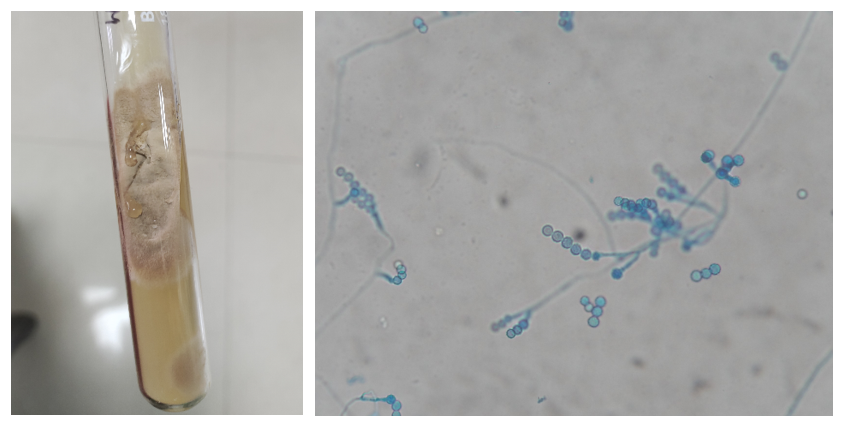
Fig. 1: Scopulariopsis brevicaulis - growth on Sabourauds’ Dextrose Agar (SDA) and slide culture Lactophenol cotton blue mount
The postoperative period was uneventful. The patient was discharged with aceclofenac-paracetamol-serratiopeptidase twice daily, montelukast-levocetirizine once daily at bedtime, xylometazoline nasal drops thrice daily, and intranasal corticosteroid spray twice daily. Patient came for follow-up after a month and later at 4 months. Follow-up period was also uneventful.
DISCUSSION
AFRS is typically a non-invasive manifestation of fungal rhinosinusitis occurring in immunocompetent hosts[4]. A key reason this report is clinically important is the uncommon etiologic agent. Scopulariopsis species are usually regarded as environmental saprophytes and are most often linked to superficial disease, while deep sinonasal disease is rare and more frequently described in immunocompromised hosts[6]. In contrast, our patient had no systemic comorbidities, normal baseline investigations, and no immunosuppressive history.
Non-invasive fungal sinusitis described by Rai S et al was observed in an immunocompetent adult male involving the maxillary sinus and responded well to surgery and itraconazole[7]. Sattler et al. described a non-invasive maxillary “fungal ball” due to S. brevicaulis in an immunocompetent 70-year-old, where surgery alone achieved good outcome despite the isolate demonstrating high MICs to multiple antifungal agents[4]. In contrast, invasive sinonasal infections attributed to Scopulariopsis have largely been confined to immunocompromised settings and tend to follow a more aggressive course. Such cases often require a combination of prompt surgical debridement and systemic antifungal therapy to achieve disease control[8, 9].
S. brevicaulis presents a constant dilemma with in vitro resistance, interpretive breakpoints lacking, clinical response not correlating well with MICs[4]. Contemporary literature suggests that no single antifungal agent has demonstrated consistent efficacy, and combination regimens with different antifungals have been explored with variable success[5, 9, 10]. It is important to understand the antifungal susceptibility in such cases, however, in the present case, complete clinical resolution was achieved with surgical excision alone, underscoring the potential adequacy of surgery in non-invasive disease. Nevertheless, certain limitations must be acknowledged. Serum IgE levels were not assessed, which could have provided additional support for the diagnosis of AFRS, and identification of Scopulariopsis brevicaulis was based solely on phenotypic characteristics without molecular confirmation.
CONCLUSION
This case broadens current understanding of sinonasal Scopulariopsis by demonstrating that S. brevicaulis can present as AFRS in an immunocompetent young adult, reinforcing the need for species-level identification. Reporting such uncommon presentations contributes to a better understanding for the role of surgery as the cornerstone of management in such non-invasive disease, while reserving systemic antifungal therapy for carefully selected cases based on host status and disease severity.
Acknowledgement
We acknowledge the contribution of our technical staff, Ms Ruby Suria and Mr Sheetal Kumar
Conflict of interest statement
The authors declare no conflict of interest.
References
1. Bent JP, Kuhn FA. Diagnosis of Allergic Fungal Sinusitis. Otolaryngology–Head and Neck Surgery. 1994; 111 (5). Available from: https://doi.org/10.1177/019459989411100508
2. Varun KMG, Kanitha MS, Rejitha IM. Fungal infection among patients with chronic rhinosinusitis who underwent endoscopic sinus surgery. International Journal of Otorhinolaryngology and Head and Neck Surgery. 2025; 11 (1). Available from: https://doi.org/10.18203/issn.2454-5929.ijohns20250114
3. Kaplan NM, Al-Dwairi RA, AlRabadi NN. Fungal keratitis due to Scopulariopsis brevicaulis and a potential promising therapeutic effect of antibacterial agents: A case report. Medicine. 2021; 100 (49). Available from: https://doi.org/10.1097/md.0000000000028203
4. Sattler L, Sabou M, Ganeval-Stoll A, Dissaux C, Candolfi E, Letscher-Bru V. Sinusitis caused by Scopulariopsis brevicaulis: Case report and review of the literature. Medical Mycology Case Reports. 2014; 5 Available from: https://doi.org/10.1016/j.mmcr.2014.05.003
5. Korvin K, Chiarruzi M, Martin A, Gromoff C, Ciobotaru V, Sasso M, et al. Endocarditis caused by Scopulariopsis brevicaulis with disseminated emboli and multiple vascular aneurysms: A case report and literature review. IDCases. 2025; 42 Available from: https://doi.org/10.1016/j.idcr.2025.e02366
6. Jabor MA, Greer DL, Amedee RG. Scopulariopsis: An Invasive Nasal Infection. American Journal of Rhinology. 1998; 12 (5). Available from: https://doi.org/10.2500/105065898780182453
7. Rai S, Tiwari R, Sandhu SV, Rajkumar Y. Hyalohyphomycosis of maxillary antrum. Journal of Oral and Maxillofacial Pathology. 2012; 16 (1). Available from: https://doi.org/10.4103/0973-029x.92996
8. Gluck O, Segal N, Yariv F, Polacheck I, Puterman M, Greenberg D, et al. Pediatric invasive sinonasal Scopulariopsis brevicaulis - a case report and literature review. International Journal of Pediatric Otorhinolaryngology. 2011; 75 (7). Available from: https://doi.org/10.1016/j.ijporl.2011.03.026
9. Ellison MD, Hung RT, Harris K, Campbell BH. Report of the first case of invasive fungal sinusitis caused by Scopulariopsis acremonium: review of scopulariopsis infections. Archives of Otolaryngology–Head & Neck Surgery. 1998; 124 (9). Available from: https://doi.org/10.1001/archotol.124.9.1014
10. Nevil V, Suma R, Arjun GM, Pooja P. Orbital apex syndrome due to scopulariopsis: a rare case report. International Journal of Advances in Medicine. 2015; Available from: https://doi.org/10.18203/2349-3933.ijam20150569
Copyright
©2026 Dhawan et al. This is an open-access journal, and articles are distributed under the terms of the Creative Commons Attribution License CC-BY 4.0. (https://creativecommons.org/licenses/by/4.0/) which permits unrestricted use, distribution, and reproduction in any medium, provided the original authors and source are credited.
Cite this article
Dhawan P, Mahajan A, Gulati N, Singla N, Gupta N, Chaudhary P, Gupta V. Allergic Fungal Rhinosinusitis Associated with Scopulariopsis brevicaulis: A Rare Case Report. Perspectives in Medical Research. 2026; 14(2):100-102 DOI: 10.47799/pimr.1402.26.44